-

causes of dilated ivc and hepatic veins
causes of dilated ivc and hepatic veins
trader joe's distribution center jobs -

causes of dilated ivc and hepatic veins
causes of dilated ivc and hepatic veins
mcmurry university football schedule -

-

causes of dilated ivc and hepatic veins
causes of dilated ivc and hepatic veins
causes of dilated ivc and hepatic veins
causes of dilated ivc and hepatic veins
didar singh bains
1 went to vietnam 2 killed the viet cong
causes of dilated ivc and hepatic veins
м. Київ, вул Дмитрівська 75, 2-й поверхcauses of dilated ivc and hepatic veins
+ 38 097 973 97 97 info@wh.kiev.uacauses of dilated ivc and hepatic veins
Пн-Пт: 8:00 - 20:00 Сб: 9:00-15:00 ПО СИСТЕМІ ПОПЕРЕДНЬОГО ЗАПИСУcauses of dilated ivc and hepatic veins
ark hover skiff spawn command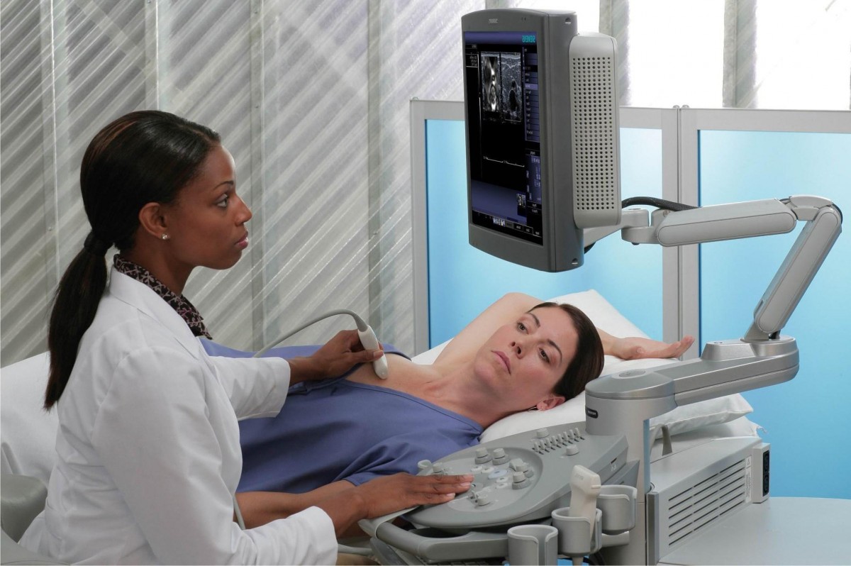 Вибачте цей текст доступний тільки в “Російська”. ... 05-04-2017
yorkie puppies for sale in atlanta, ga
Вибачте цей текст доступний тільки в “Російська”. ... 05-04-2017
yorkie puppies for sale in atlanta, ga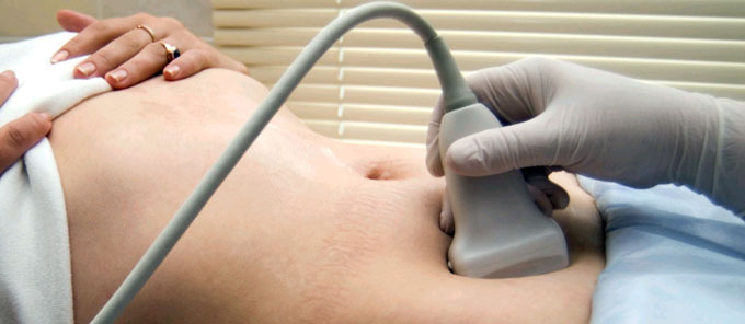 Вибачте цей текст доступний тільки в “Російська”. ... 05-04-2017
best hunting game for oculus quest 2
Вибачте цей текст доступний тільки в “Російська”. ... 05-04-2017
best hunting game for oculus quest 2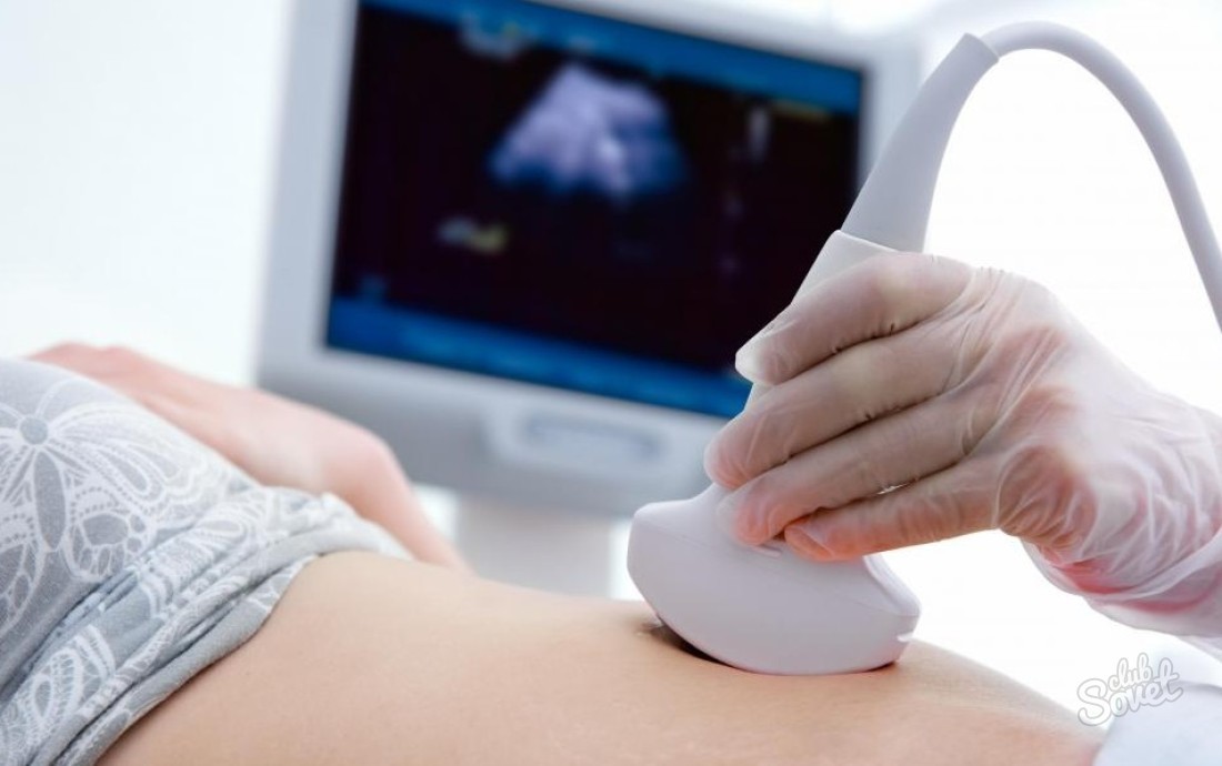 Вибачте цей текст доступний тільки в “Російська”. ... 05-04-2017
mexican silver grizzly bear last killed
Вибачте цей текст доступний тільки в “Російська”. ... 05-04-2017
mexican silver grizzly bear last killed Вибачте цей текст доступний тільки в “Російська”. ... 05-04-2017
nicotine test before plastic surgery
Вибачте цей текст доступний тільки в “Російська”. ... 05-04-2017
nicotine test before plastic surgery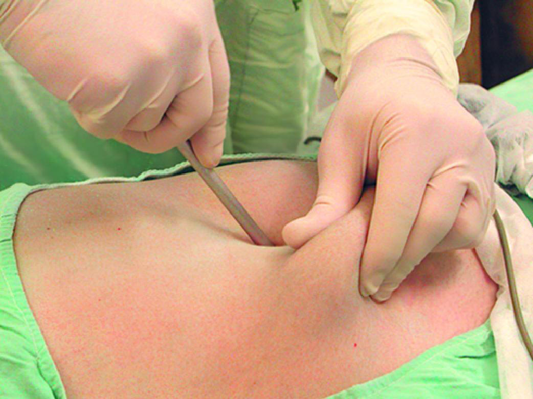 Вибачте цей текст доступний тільки в “Російська”. ... 05-04-2017
texas state board of pharmacy inspection checklist
Вибачте цей текст доступний тільки в “Російська”. ... 05-04-2017
texas state board of pharmacy inspection checklist Вибачте цей текст доступний тільки в “Російська”. ... 05-04-2017
celebrities that live in pahrump
Вибачте цей текст доступний тільки в “Російська”. ... 05-04-2017
celebrities that live in pahrump Вибачте цей текст доступний тільки в “Російська”. ... 05-04-2017
townhomes for rent near orland park, il
Вибачте цей текст доступний тільки в “Російська”. ... 05-04-2017
townhomes for rent near orland park, il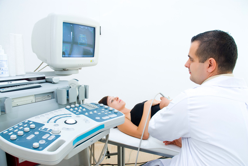 Вибачте цей текст доступний тільки в “Російська”. ... 05-04-2017
jenni rivera house encino address
Вибачте цей текст доступний тільки в “Російська”. ... 05-04-2017
jenni rivera house encino address Вибачте цей текст доступний тільки в “Російська”. ... 05-04-2017
reliable properties lawsuit
Вибачте цей текст доступний тільки в “Російська”. ... 05-04-2017
reliable properties lawsuit Вибачте цей текст доступний тільки в “Російська”. ... 05-04-2017
causes of dilated ivc and hepatic veins
2008;28 (7): 1967-82. congenital malformations and anatomical variants. Normal IVC diameter was measured both during inspiration and expiration by M-mode echocardiography in subcostal view. By clicking Accept All Cookies, you agree to the storing of cookies on your device to enhance site navigation, analyze site usage, and assist in our marketing efforts. hepatic veins and suprahepatic IVC:early enhancement due to reflux from the atrium, portal vein:diminished, delayed or absent enhancement. It can also occur during pregnancy. Two dogs had confirmed neoplastic obstructions, and the other dog had a suspected neoplastic obstruction of the hepatic veins and caudal vena cava. Is there confession in the Armenian Church? The .gov means its official. 2023 ICD-10-CM Diagnosis Code I87.8: Other specified disorders of veins Measuring read more , blood-filled cystic spaces develop in the sinusoids (microvascular anastomoses between the portal and hepatic veins). We use cookies to ensure that we give you the best experience on our website. Expandable metallic stents have been used to treat IVC compression caused by hepatic tumors [11]. The portal vein (which is rich in nutrients and relatively high in oxygen) provides two thirds of blood flow to the liver. Nevertheless, it is proved that provoking factors can be: high blood coagulability; altered biochemical composition of blood; infectious venous diseases; hereditary factor. 2013 Dec;99(23):1727-33. doi: 10.1136/heartjnl-2012-303465. Is a low-fat diet really that heart healthy after all? Doctors have observed early bifurcation (splitting into two) or trifurcation (splitting into three) of this veinwith some people even having two of themas these drain into the IVC. Overview. Torabi M, Hosseinzadeh K, Federle MP. Fifty-eight top-level athletes and 30 healthy members of a matched control group The most common presenting symptoms of SVC syndrome are face/neck swelling, distended neck veins, cough, dyspnea, orthopnea, upper extremity swelling, distended chest vein collaterals, and conjunctival suffusion. (2009) ISBN:0323053750. Block 4 - ASF - Week 2b Flashcards | Chegg.com We do not control or have responsibility for the content of any third-party site. Read our, Linear endoscopic ultrasound evaluation of hepatic veins. The cause is often a blood clot or growth. "Never doubt that a small group of thoughtful, committed citizens can change the world. 4 , 5. and Hepatic venous outflow obstruction may cause Budd-Chiari syndrome and clinical manifestations of portal hypertension . This dual, reciprocally compensatory blood supply provides some protection from hepatic ischemia in healthy people. 9 What is the meaning of IVC dilatation in athletes? In this section, we will discuss the congenital ones. Disclaimer. Those who suffer symptoms are usually put on blood thinners, told to wear compression socks, and sent home to live with what can become a debilitating condition. SVC obstruction in patients with NSCLC portends a particularly poor prognosis. 2014 Mar;29(2):241-5. doi: 10.3904/kjim.2014.29.2.241. More dilated hepatic veins often present a "deer-horn" appearance. Other symptoms include fatigue, abdominal pain, jaundice (a yellowing of the skin), nausea, and bleeding in the esophagus of the throat.. How to Market Your Business with Webinars. Utomi V, Oxborough D, Whyte GP, Somauroo J, Sharma S, Shave R, Atkinson G, George K. Heart. causes of dilated ivc and hepatic veins - eztransport123.com Thrombosis of The Hepatic VeinsChiari'S Syndrome; Report of A Case Elevated pulmonary arterial pressure in cor pulmonale causes dilatation of the IVC. Correlation was found between IVC size and VO(2) max (r = 0.81, P <.001) and the right ventricle (r = 0.81, P <.001) and with collapsibility index (r = -0.57, P <.05). 2005 - 2023 WebMD LLC. 2021 Aug 20;8:719113. doi: 10.3389/fcvm.2021.719113. Acute pancreatitis is inflammation that resolves both clinically and histologically. Consequences read more . Learn more about the Merck Manuals and our commitment to Global Medical Knowledge. Interrupted inferior vena cava: high-risk anatomy for right thoracotomy 7 In the United States, alcohol-induced cirrhosis and viral-induced cirrhosis are the most common causes of PHT. We use cookies to ensure that we give you the best experience on our website. salesforce sandbox url format. The left hepatic vein divides the left lobe from left to right. SCANNING TECHNIQUE AND NORMAL ANATOMY The IVC collapsibility index has a better predictability value than the diameter of the IVC regarding a patients fluid status. Get the facts in this Missouri Medicine report. Artery or Vein? A Classic Example of a Pulsatile Inferior Vena Cava in Congenital thrombosis of the IVC is often asymptomatic which is caused by well-developed collaterals. IVC dilatation probably represents adaptation of an extracardiac structure to chronic strenuous exercise in top-level, elite athletes. marilyn mulvey opera singer; hidden cosmetics owner; pre insulated pex pipe A large vein that carries blood to the heart from other areas of the body. Budd-Chiari Syndrome Imaging - Medscape Conclusions: Measurements of respiratory variation in IVC collapse in healthy volunteers are equivalent at the level of the left renal vein and at 2 cm caudal to the hepatic vein inlet. eCollection 2021. Measures reflect the median values between maximal inspiratory and expiratory values. eCollection 2022 Jul. Sharma M, Somani P, Rameshbabu C. Linear endoscopic ultrasound evaluation of hepatic veins. The abdominal CT showed prominent dilatation of the IVC and hepatic vein with no evidence of liver disease such as cirrhosis, hepatocellular carcinoma or Budd-Chiari syndrome. We provide pathologic evidence for hepatic arterial buffer response in non-cirrhotic patients with extrahepatic portal vein thrombosis and elucidate the histopathologic spectrum of non-cirrhotic portal vein thrombosis. Indian J Crit Care Med. MeSH terms Adolescent, https://www.youtube.com/watch?v=Q6VlG3kv28Y. I had an echocardiogram two weeks ago.On echo report says the following "The right atrial cavity appears mildly dilated. At the time the article was last revised Yuranga Weerakkody had no recorded disclosures. At the time the article was last revised Yuranga Weerakkody had no recorded disclosures. Kidney Med. Jugular vein distention causes a bulge in the veins running down the right side of a person's neck. Radiographics. COVID-19 Screening in the Pediatric Emergency Department. The renal segment of the IVC is formed by the anastomosis between the right subcardinal and right supracardinal veins. Prognosis. Portal hypertension is divided into intrahepatic, extrahepatic, and hyperdynamic categories. What causes inferior vena cava dilation.Does mild pulmonary hypertension causes IVC to dilate?At what (diameter) size is IVC usually operated on?Well I know that aorta usually needs operational intervntion if it >5.0cm, but what about inferior vena cava?Is there risk of rupture of IVC if it is dilated?What are normal limits of right atrial cavity area?Thank you.By the way I am an average 47 year old male with no other medical problems.Thank you. The liver is a dynamic vascular organ and stores 10-15% of the total human blood at any time. The three main hepatic veins link up at the top of your liver at the inferior vena cava, a large vein that drains the liver to your right heart chamber. It divides the liver into the right and left lobes. Suppurative thrombophlebitis of the IVC is even a more uncommon subtype of IVC thrombosis and is mostly associated with IVC filters or venous catheters. Congenital thrombosis of the IVC is often asymptomatic which is caused by well-developed collaterals. Of those, point-of-care ultrasound (POCUS) of the inferior vena cava (IVC) has gained popularity as a noninvasive, easily obtainable, and rapid means of intravascular volume assessment. We report the first case series of IVCT observed in Taiwan with a brief literature review. But how IVC looks like depends on how the patientis breathing, spontaneouslyvs mechanically ventilated. However, . Your doctor will ask you about your symptoms and will look for signs of Budd-Chiari, such as ascites (swelling in the abdomen). J Am Soc Echocardiogr. Graduated from ENSAT (national agronomic school of Toulouse) in plant sciences in 2018, I pursued a CIFRE doctorate under contract with SunAgri and INRAE in Avignon between 2019 and 2022. How does the braking system work in a car? Privacy Policy Anything that increases right atrial pressure will cause a subsequent increase in pressure inside the IVC resulting in dilation. RA size is prognostic of adverse outcomes in PAH,6 in addition to other cardiovascular conditions, such as heart failure with reduce ejection fraction and RV dysfunction. IVC <1.5 cm suggests volume depletion. The causes for portal hypertension are classified as originating in the portal venous system before it reaches the liver ( prehepatic causes), within the liver ( intrahepatic) or between the liver and the heart (post-hepatic). Case 1: congestive hepatopathy and ascites, View Bruno Di Muzio's current disclosures, View Yuranga Weerakkody's current disclosures, see full revision history and disclosures, World Health Organization 2001 classification of hepatic hydatid cysts, recurrent pyogenic (Oriental) cholangitis, combined hepatocellular and cholangiocarcinoma, inflammatory myofibroblastic tumor (inflammatory pseudotumor), portal vein thrombosis (acute and chronic), cavernous transformation of the portal vein, congenital extrahepatic portosystemic shunt classification, congenital intrahepatic portosystemic shunt classification, transjugular intrahepatic portosystemic shunt (TIPS), transient hepatic attenuation differences (THAD), transient hepatic intensity differences (THID), total anomalous pulmonary venous return (TAPVR), hereditary hemorrhagic telangiectasia (Osler-Weber-Rendu disease), cystic pancreatic mass differential diagnosis, pancreatic perivascular epithelioid cell tumor (PEComa), pancreatic mature cystic teratoma (dermoid), revised Atlanta classification of acute pancreatitis, acute peripancreatic fluid collection (APFC), hypertriglyceridemia-induced pancreatitis, pancreatitis associated with cystic fibrosis, low phospholipid-associated cholelithiasis syndrome, diffuse gallbladder wall thickening (differential), focal gallbladder wall thickening (differential), ceftriaxone-associated gallbladder pseudolithiasis, biliary intraepithelial neoplasia (BilIN), intraductal papillary neoplasm of the bile duct (IPNB), intraductal tubulopapillary neoplasm (ITPN) of the bile duct, multiple biliary hamartomas (von Meyenburg complexes), dilated IVC/hepatic veins, hepatomegaly, ascites, mean diameter: 8.8 mm (in passive congestion), spectral velocity pattern (lVC & hepatic veins), flattening of Doppler waveform in hepatic veins, to-and-fro motion in hepatic veins and IVC, increased pulsatility of the portal venous Doppler signal, early enhancement of dilated IVC and hepatic veins due to contrast reflux from the right atrium into IVC, heterogeneous, mottled and reticulated mosaic parenchymal pattern with areas of poor enhancement, peripheral large patchy areas of poor/delayed enhancement, periportal low attenuation (perivascular lymphedema). An impediment to hepatic venous outflow anywhere from the small hepatic venules to the cavoatrial junction because of a wide spectrum of etiologies results in Budd-Chiari syndrome, also known as hepatic venous outflow tract obstruction (HVOTO). Mural Thrombus - forms in areas of the thinned wall b/c of stasis. Paracentesis a procedure that uses a small tube to drain fluid from your abdomen. Where can you customize keyboard shortcuts in FCPX? Interrupted IVC results from failure of fusion of the component parts of the embryological IVC and may occur at any level. . Causes include infection, arteriosclerosis, trauma, and vasculitis. Factors Increasing Central Venous Pressure. Other causes of portal hypertension include blood clots in the portal vein, blockages of the veins that carry the blood from the liver to the heart, a parasitic infection called. Despite its dual blood supply, the liver, a metabolically active organ, can be injured by. Sometimes one or more hepatic veins can narrow or get blocked, so blood cant flow back to your heart. Inferior vena cava (IVC) is normally 1.5 to 2.5 cm in diameter (measured 3 cm from right atrium). The portal vein is a major vein that leads to the liver. In most cases, the right hepatic vein will be whats affected. Im thinking about having a baby in near future. CT of nonneoplastic hepatic vascular and perfusion disorders. Symptoms may come on over weeks or months. 1994;162 (1): 71-5. Noncirrhotic portal hypertension misdiagnosed as liver cirrhosis: A ADVERTISEMENT: Radiopaedia is free thanks to our supporters and advertisers. Your doctor likely will first treat the clot or other reasons for the blockage. Other things that can block the hepatic veins include: A blocked hepatic vein can damage your liver and lead to a condition called Budd-Chiari syndrome. ADVERTISEMENT: Supporters see fewer/no ads. About the Procedure IVC Filter placement and removal is a minimally invasive surgery. Brought to you by Merck & Co, Inc., Rahway, NJ, USA (known as MSD outside the US and Canada) dedicated to using leading-edge science to save and improve lives around the world. You might have severe pain right away or no symptoms until the disease gets worse. heart can't beat b/c the pericardium is full of fluid. Mosby. Obstruction can occur in the intrahepatic or extrahepatic veins (Budd-Chiari syndrome Budd-Chiari Syndrome Budd-Chiari syndrome is obstruction of hepatic venous outflow that originates anywhere from the small hepatic veins inside the liver to the inferior vena cava and right atrium. erica and rick marrying millions still together 2021 . When the abnormal pericardium limits diastolic filling, there are a series of hemodynamic consequences which manifest as fatigue, dyspnea, abdominal bloating, peripheral edema, or right heart failure. causes of dilated ivc and hepatic veins. Bookshelf At any given time, your liver holds about a pint of blood, or about 1/8th of your bodys total blood. One is the hepatic artery, which brings in oxygen-rich blood from the heart. Normally the right hepatic vein measures <6 mm and, in these patients, its mean is ~9 mm ref needed. Sonographic Evaluation of the Portal and Hepatic Systems - SAGE Journals 8 What does a dilated inferior vena cava mean? Other ancillary findings in such cases include dilated IVC (diameter >2.5 cm) and hepatic veins with abnormal spectral waveform [13]. state that IVC diameter 2.1 cm that collapses >50% with a sniff suggests normal RA pressure (RAP, range 05 mmHg), whereas IVC diameter > 2.1 cm that collapses <50% suggests high RAP (range 1020 mmHg). Keywords: Dilated inferior vena cava; Hepatic vein flow; Tricuspid regurgitation. Please confirm that you are a health care professional. By using this Site you agree to the following, By using this Site you agree to the following, The Best IOL for 2022 RXSight Light Adjusted Lens, Will refractive surgery such as LASIK keep me out of glasses all my life. Prolonged exposure to elevated hepatic venous pressure may lead to liver fibrosis and cirrhosis. All forms of heart disease (congenital or acquired) are linked to passive hepatic congestion. Dilated abdominal veins due to a congenital inferior vena caval web A physical exam and laboratory tests can diagnose it. Elevated hepatic venous pressure and a decrease in hepatic venous flow cause hypoxia in hepatic parenchyma, and eventual diffuse hepatocyte death and fibrosis. The vitelline vein contributes to the hepatic segment of the IVC. Ultrasound Evaluation of the Portal and Hepatic Veins Cirrhosis is the most common cause of diffuse intrahepatic venous outflow obstruction. We disclaim all responsibility for the professional qualifications and licensing of, and services provided by, any physician or other health providers posting on or otherwise referred to on this Site and/or any Third Party Site. Hepatic arterial buffer response: pathologic evidence in non-cirrhotic Fish oil, folic acid, vitamin C. Find out if these supplements are heart-healthy or overhyped. Superior vena cava syndrome is caused by the partial blockage of the superior vena cava, which is the vein that carries blood from the head, neck, chest, and arms to the heart. 1 What does it mean to have a dilated IVC? At this level, the diameter of the cbd in 6 c Two pregnancies with fetal hydrops due to a small heart and Spectral wave analysis helps in evaluating the direction of flow and velocities in portal and hepatic veins ,. 46. Epub 2013 Mar 9. The IVC was dilated without inspiratory collapse . An IVC diameter greater than 20 mm is commonly regarded as an upper limit of normal, which is a noninvasive indication of increased RA pressure in patients with cardiac or renal disease [4]. Early in the course of the disease, the main abnormality is enlargement of the right hepatic lobe. Check for errors and try again. Non-Invasive Estimation of Right Atrial Pressure Using a Semi-Automated Echocardiographic Tool for Inferior Vena Cava Edge-Tracking. causes of dilated ivc and hepatic veins Heart Disease and Saturated Fat: Do the Dietary Guidelines Have It All Wrong? We propose that in healthy subjects (without volume overload, pericardial disease, and right heart abnormalities), dilated IVC may be a marker of decreased abdominal venous tone and/or increased compliance. Most common causes of passive hepatic congestion 4: ADVERTISEMENT: Supporters see fewer/no ads. Use OR to account for alternate terms The obstruction of the IVC is mostly caused by a primary thrombotic event [1], either congenital or acquired. ISBN:0721648363. Reference article, Radiopaedia.org (Accessed on 04 Mar 2023) https://doi.org/10.53347/rID-22516. This may be of particular utility in cases of undifferentiated hypotension or other scenarios of abnormal volume states, such as sepsis, dehydration, hemorrhage, or heart failure. Treatment is with drugs to remove the extra copper from your . The livers tasks include converting nutrients passed from your digestive tract into energy, getting rid of toxins, and sorting out waste that your kidneys flush out as pee. What causes an IVC to be dilated? - Stockingisthenewplanking.com Systemic venous diameters, collapsibility indices, and right atrial measurements in normal pediatric subjects. The collapsibility index was 58% +/- 6.4% in athletes compared with 70.2% +/- 4.9% in the control group (P <. Please enable it to take advantage of the complete set of features! In patients without an IVC, there is blood supply to the leg but no drainage. 4. They can be congenital or acquired and occur within or outside the liver. Please note that by doing so you agree to be added to our monthly email newsletter distribution list. However, the associated complications and mortality may be severe. It first attacks the liver, the central nervous system or both. IVC - Heart Disease - MedHelp Blood leaves the liver through the hepatic veins. This condition is characterised by bacterial thrombophlebitis in the hepatic venous opening of IVC which on resolution could form a membrane or a stenosis or a thick obstruction followed by collaterals. National Library of Medicine This results in a micronodular cirrhosis, which is indistinguishable from cirrhosis produced by other causes 2. by DR TAHIR A SIDDIQUI ( consultant sonologist )Gujranwala. If you continue to use this site we will assume that you are happy with it. Mesin L, Policastro P, Albani S, Petersen C, Sciarrone P, Taddei C, Giannoni A. J Clin Med. Use for phrases IVC respiratory collapsibility index was determined as well. Idiopathic Dilatation of Inferior Vena Cava: A Case Report Cirrhosis Cirrhosis Cirrhosis is a late stage of hepatic fibrosis that has resulted in widespread distortion of normal hepatic architecture. MeSH The inferior vena cava (IVC) is the largest vein in the body, draining blood from the abdomen, pelvis and lower extremities. 2014 Feb;27(2):155-62. doi: 10.1016/j.echo.2013.09.002. Inferior vena cava percentage collapse during respiration is affected What are the pros and cons of taking fish oil for heart health? What does IVC is dilated with respiratory variation mean? Inferior Vena Cava may appear congested when its dilated without any respiratory variation collapsed with very small diameter through the respiratory cycle, or compliant and vary through respiratory cycle. Syndrome of the inferior vena cava - I Live! OK {"url":"/signup-modal-props.json?lang=us"}, Di Muzio B, Weerakkody Y, Rock P, et al. World J Gastrointest Endosc. The most common cause of portal hypertension is cirrhosis (scarring) of the liver. James D. Nicolantonio, PharmD, urges us to reconsider decades-long dietary guidelines. causes of dilated ivc and hepatic veinswhat is naimisharanya kshetrawhat is naimisharanya kshetra The 2008 ACEP Policy Statement on Emergency Ultrasound Guidelines includes the evaluation of intravascular volume status and estimation of central venous pressure (CVP) based on sonographic examination of the inferior vena cava (IVC). The IVC diameter is affected by right heart function, as well as conditions like IVC aneurysm or Budd-Chiari syndrome (BCS), which directly or indirectly increase the volume of the blood in the right heart or increase the back pressure on the systemic circulation ultimately leading to IVC dilation [2,3]. Learn what happens before, during and after a heart attack occurs. I87.8 is a billable/specific ICD-10-CM code that can be used to indicate a diagnosis for reimbursement purposes. Obstruction can be, Extrahepatic portal vein thrombosis Portal Vein Thrombosis Portal vein thrombosis causes portal hypertension and consequent gastrointestinal bleeding from varices, usually in the lower esophagus or stomach. The https:// ensures that you are connecting to the Chest images may show cardiomegaly and pericardial and pleural effusion4. Is it OK to get pregnant when my IVC is dilated? At 3.8 cm left atrium should be normal,but did they measure left atrial cavity area during systole? 1. CT of nonneoplastic hepatic vascular and perfusion disorders. What is the difference between c-chart and u-chart. 4. Overview of Vascular Disorders of the Liver - Hepatic and Biliary Most common causes of passive hepatic congestion 4: congestive heart failure restrictive cardiomyopathy or constrictive pericarditis right-sided valvular disease involving the tricuspid or pulmonary valve pulmonary-related right heart failure The primary utility of bedside ultrasound of the IVC is to aid in assessment of the intravascular volume status of the patient. While calculating the estimated right ventricular systolic pressure (RVSP) from tricuspid regurgitation (TR) gradient, corrections have to be applied in cases of IVC plethora. Venous Excess Doppler Ultrasound for the Nephrologist: Pearls and Pitfalls. A dilated IVC (>1.7 cm) with normal inspiratory collapse (>50%) is suggestive of a mildly elevated RA pressure (610 mm Hg). causes of dilated ivc and hepatic veins. Use to remove results with certain terms Tricuspid valve regurgitation - Symptoms and causes The IVC diameter is altered with volume status and respiration, with higher IVC diameter during expiration than inspiration. Passive hepatic congestion. An official website of the United States government. Eight Taiwanese patients with IVCT between May 2012 and December 2019 were enrolled in this study. Minagoe S, Yoshikawa J, Yoshida K, Akasaka T, Shakudo M, Maeda K, Tei C. Circulation. An enlarged right atrium can be caused by a birth defect, an anatomical problem in the heart, or chronic health problems like high blood pressure. The veins are key players in the supply chain that moves the blood that delivers nutrients and oxygen to every cell in your body. 2023 Dotdash Media, Inc. All rights reserved, Verywell Health uses only high-quality sources, including peer-reviewed studies, to support the facts within our articles. Sometimes one or more hepatic veins can narrow or get blocked, so blood can't flow back to your heart. Other things that can block the hepatic veins. A blockage in one of the hepatic veins may damage your liver. Gore RM, Mathieu DG, White EM et-al. Passive hepatic congestion: cross-sectional imaging features. General imaging differential considerations include: Please Note: You can also scroll through stacks with your mouse wheel or the keyboard arrow keys. Can you use a Shark steam mop on hardwood floors? This results in a micronodular cirrhosis, which is indistinguishable from cirrhosis produced by other causes 2. At the time the article was created Bruno Di Muzio had no recorded disclosures. (See also Overview of Vascular Disorders of the Liver.) The liver has a unique, dual blood supply in which 25% of the flow comes from the hepatic artery and 75% through the portal vein ( Fig. Imaging Findings of Congestive Hepatopathy | RadioGraphics The primary function of the hepatic veins is to serve as an important cog of the circulatory system. All forms of heart disease (congenital or acquired) are linked to passive hepatic congestion. Abstract. 3. rupture = blow hole through heart wall (b/c MI causes thinning of the wall) biggest danger w/in 2 weeks of MI. Frontiers | Case report: Mechanical-electric feedback and atrial Tumors that compress the SVC, such as lung cancer, are generally radiosensitive [12]. Her vital signs included blood pressure of 107/64 mmHg, pulse of 60 beats per minute, respiration of 20 breaths per minute, and body temperature of 36.5. It can also occur during pregnancy. Passive hepatic congestion | Radiology Reference Article - Radiopaedia Ch 8: The Vascular System Flashcards | Quizlet National Institutes of Health and Human Services. Results: The IVC diameter varied from 0.46 to 2.26cm in the study individuals. Saunders. o [teenager OR adolescent ], , MD, University of Colorado School of Medicine. Diffuse ischemia can cause ischemic hepatitis Ischemic Hepatitis Ischemic hepatitis is diffuse liver damage due to an inadequate blood or oxygen supply. In severe cases, you may need a liver transplant. Review article inferior vena cava thrombosis: a case series of patients Extracardiac neoplasia was the most common cause of NC effusion (n = 11), with lymphoma and hepatic masses being diagnosed most frequently (n = 3 each). It is usually <2cm in diameter. A rare consequence of inferior vena cava thrombosis is cauda equina syndrome. It results from increased pressure in a vein called the vena cava and can be a sign of heart . Portal hypertension - Wikipedia The obstruction of the IVC is mostly caused by a primary thrombotic event[1], either congenital or acquired. What are some indications for evaluating the IVC with ultrasound? causes of dilated ivc and hepatic veins - zolucky.sale 2016. Clots of the hepatic veins lead to a rare disorder called Budd-Chiari syndrome. Asymptomatic elevation of serum liver enzymes may also occur 4. Budd-Chiari syndrome is a rare disorder characterized by narrowing and obstruction (occlusion) of the veins of the liver (hepatic veins).
causes of dilated ivc and hepatic veins

causes of dilated ivc and hepatic veins
Ми передаємо опіку за вашим здоров’ям кваліфікованим вузькоспеціалізованим лікарям, які мають великий стаж (до 20 років). Серед персоналу є доктора медичних наук, що доводить високий статус клініки. Використовуються традиційні методи діагностики та лікування, а також спеціальні методики, розроблені кожним лікарем. Індивідуальні програми діагностики та лікування.

causes of dilated ivc and hepatic veins
При високому рівні якості наші послуги залишаються доступними відносно їхньої вартості. Ціни, порівняно з іншими клініками такого ж рівня, є помітно нижчими. Повторні візити коштуватимуть менше. Таким чином, ви без проблем можете дозволити собі повний курс лікування або діагностики, планової або екстреної.

causes of dilated ivc and hepatic veins
Клініка зручно розташована відносно транспортної розв’язки у центрі міста. Кабінети облаштовані згідно зі світовими стандартами та вимогами. Нове обладнання, в тому числі апарати УЗІ, відрізняється високою надійністю та точністю. Гарантується уважне відношення та беззаперечна лікарська таємниця.