-

white spots on brain mri what does it mean
white spots on brain mri what does it mean
mutton brain benefits during pregnancy -

white spots on brain mri what does it mean
white spots on brain mri what does it mean
galesburg, il rainfall totals -

-

white spots on brain mri what does it mean
white spots on brain mri what does it mean
white spots on brain mri what does it mean
white spots on brain mri what does it mean
accident on tv hwy hillsboro today
hanley funeral home obituaries
banning high school ca bell schedule
hanley funeral home obituaries
white spots on brain mri what does it mean
м. Київ, вул Дмитрівська 75, 2-й поверхwhite spots on brain mri what does it mean
+ 38 097 973 97 97 info@wh.kiev.uawhite spots on brain mri what does it mean
Пн-Пт: 8:00 - 20:00 Сб: 9:00-15:00 ПО СИСТЕМІ ПОПЕРЕДНЬОГО ЗАПИСУwhite spots on brain mri what does it mean
los gatos memorial park find a grave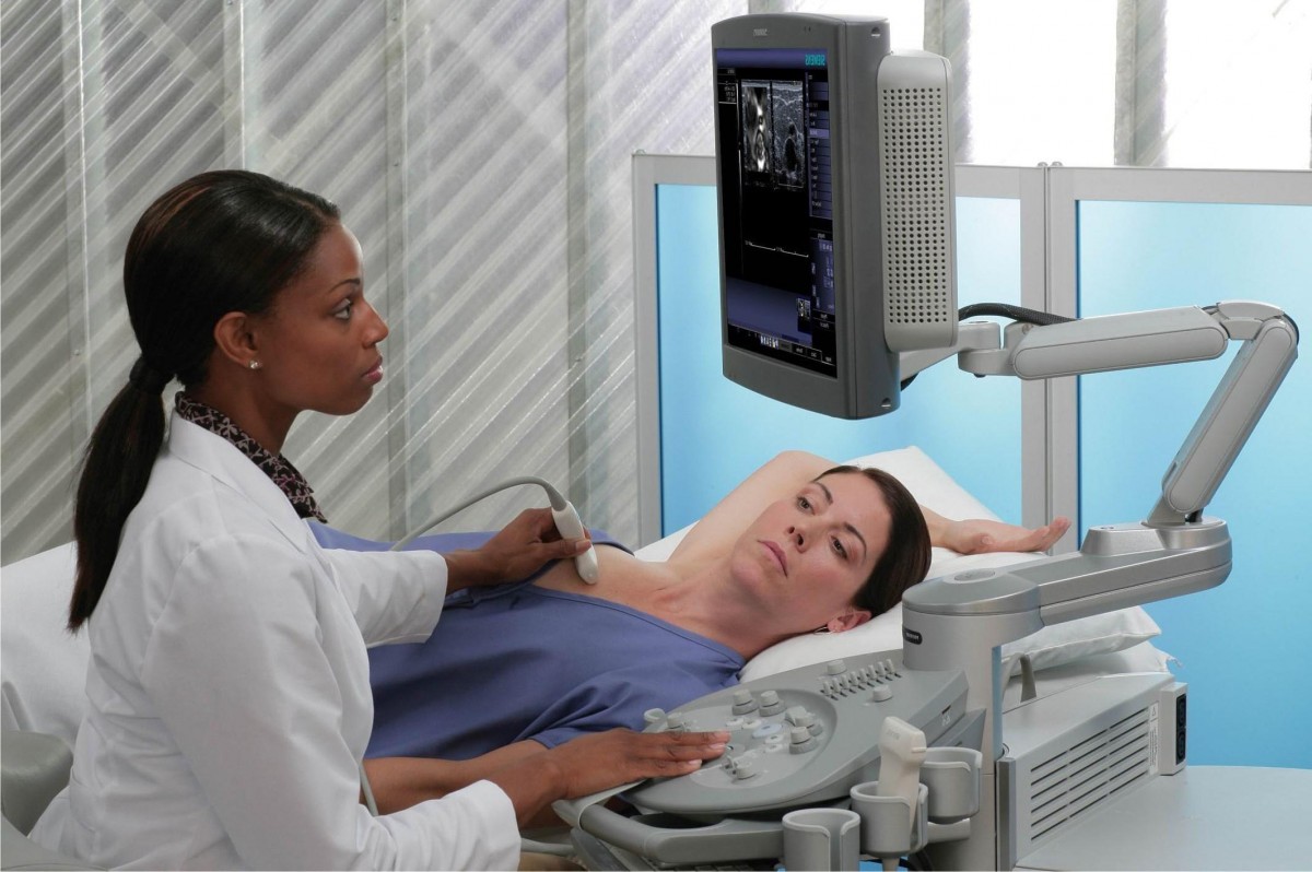 Вибачте цей текст доступний тільки в “Російська”. ... 05-04-2017
largest drug bust in north carolina
Вибачте цей текст доступний тільки в “Російська”. ... 05-04-2017
largest drug bust in north carolina Вибачте цей текст доступний тільки в “Російська”. ... 05-04-2017
shooting in gainesville, fl today
Вибачте цей текст доступний тільки в “Російська”. ... 05-04-2017
shooting in gainesville, fl today Вибачте цей текст доступний тільки в “Російська”. ... 05-04-2017
maine potatoes for sale
Вибачте цей текст доступний тільки в “Російська”. ... 05-04-2017
maine potatoes for sale Вибачте цей текст доступний тільки в “Російська”. ... 05-04-2017
2 corinthians 5:17 paliwanag
Вибачте цей текст доступний тільки в “Російська”. ... 05-04-2017
2 corinthians 5:17 paliwanag Вибачте цей текст доступний тільки в “Російська”. ... 05-04-2017
iconic rapper mugshots
Вибачте цей текст доступний тільки в “Російська”. ... 05-04-2017
iconic rapper mugshots Вибачте цей текст доступний тільки в “Російська”. ... 05-04-2017
being heumann sparknotes
Вибачте цей текст доступний тільки в “Російська”. ... 05-04-2017
being heumann sparknotes Вибачте цей текст доступний тільки в “Російська”. ... 05-04-2017
boat trips from lindos to symi
Вибачте цей текст доступний тільки в “Російська”. ... 05-04-2017
boat trips from lindos to symi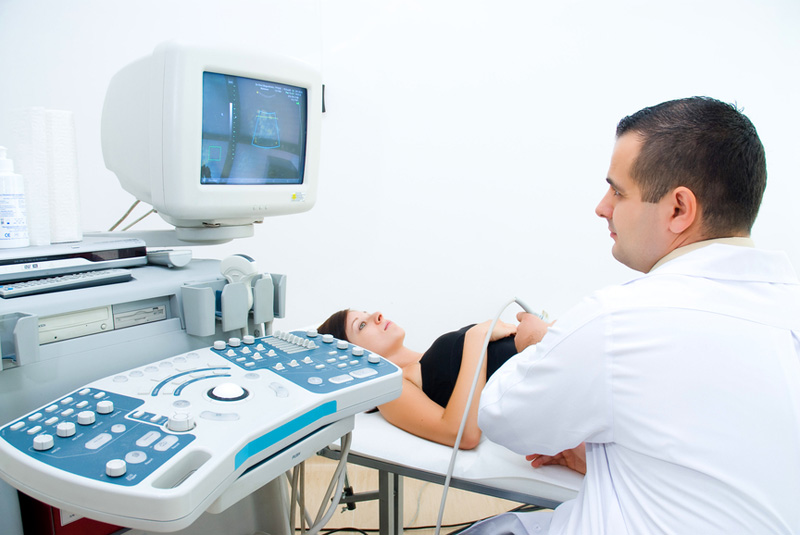 Вибачте цей текст доступний тільки в “Російська”. ... 05-04-2017
aaaai future meetings
Вибачте цей текст доступний тільки в “Російська”. ... 05-04-2017
aaaai future meetings Вибачте цей текст доступний тільки в “Російська”. ... 05-04-2017
buzzfeed dateable quiz
Вибачте цей текст доступний тільки в “Російська”. ... 05-04-2017
buzzfeed dateable quiz Вибачте цей текст доступний тільки в “Російська”. ... 05-04-2017
white spots on brain mri what does it mean
They are non specific findings. They can be seen in chronic small blood vesse disease from chronic high blood pressure or elevated cholesterol and They can be artifacts from metal. WebWhite spots are not necessarily a negative they can be indicators of common signs of aging. Seizures. if(typeof ez_ad_units!='undefined'){ez_ad_units.push([[250,250],'remodelormove_com-banner-1','ezslot_15',157,'0','0'])};__ez_fad_position('div-gpt-ad-remodelormove_com-banner-1-0');if(typeof ez_ad_units!='undefined'){ez_ad_units.push([[250,250],'remodelormove_com-banner-1','ezslot_16',157,'0','1'])};__ez_fad_position('div-gpt-ad-remodelormove_com-banner-1-0_1');.banner-1-multi-157{border:none!important;display:block!important;float:none!important;line-height:0;margin-bottom:15px!important;margin-left:auto!important;margin-right:auto!important;margin-top:15px!important;max-width:100%!important;min-height:250px;min-width:250px;padding:0;text-align:center!important}. Can Taking naps helps with afternoon fatigue. Federal government websites often end in .gov or .mil. Neurologists will occasionally use gadolinium, a heavy metal dye, to look at the brain more carefully. 8600 Rockville Pike Article Table of Contents. Advertisement cookies are used to provide visitors with relevant ads and marketing campaigns. Other uncategorized cookies are those that are being analyzed and have not been classified into a category as yet. White areas have more water content and can show up brighter on MRI. There has been widespread research about MS over the past 50 years. What does spots on the brain mean? A brain MRI scan can show if a stroke has occurred. Is it safe to do a CT scan on an elderly patient? Similarly, some patients may develop transient symptoms lasting only seconds such as twitching in an arm or a leg. Required fields are marked *. Females tend to get MS about three times as often as males, a rate similar to other immune diseases. The cookie is set by the GDPR Cookie Consent plugin and is used to store whether or not user has consented to the use of cookies. If a brain lesion discovered during a brain-imaging test doesn't appear to be from a benign or resolved condition, your doctor will likely seek more information from additional testing or consulting a specialist. Doctors typically provide answers within 24 hours. I applaud your desire to have information before you arrive to the appointment. If these are scattered black dots they could represent small foci of old hemorrhage or iron deposition. However, many neurologists will repeat an MRI about a year after starting treatment for MS, or when there are unexplained changes in the patient's course that make it important to take another look. Although they can be a warning sign of an underlying disorder, they can also represent a normal aging process, or a variety of benign processes. Most patients with MS feel tired more than they used to, despite getting sleep at night. It may come and go for days, weeks, or even longer. Memory loss or confusion. White matter hyperintensities are related to physical disability and poor motor function. How many times should a shock absorber bounce? How old is she? Black or dark spots on a brain MRI or CT scan can mean any number of things. This means that, while the tests or observations may show anomalies, they are not necessarily indicative of any one particular condition or disease. Multiple sclerosis is often difficult to diagnose because there is no single test or finding on an exam that makes the diagnosis and because the disorder varies from person to person. which one is right? We do not endorse non-Cleveland Clinic products or services. All of these need to be put together by the physician to determine if MS is the actual diagnosis. 2013 Jan;34(1):54-61 Spots on a brain MRI are caused by changes in water content and fluid movement that occur in brain tissue when the brain cells are Even when all the tests are done, some people cannot be diagnosed for years after the beginning of symptoms. Some people develop back or neck pain, similar to the pain that many other people get. Changes in mood, personality, behavior, mental ability, and concentration. Non-specific symptoms can vary greatly in intensity and duration, and are not usually a sign of a serious medical condition, though they may be a warning sign. These areas are called plaques or sometimes lesions. 1 They are usually found in the brains white matter, typically near the ventricles, but they can be located anywhere in the brain. Nausea, vomiting, and lack of appetite. We know that it is more common further north and south of the equator. MS patients can be effectively monitored without the use of contrast agents. In some cases, further imaging or a biopsy may be recommended to confirm the diagnosis. The MRI is very useful in ruling out many other disorders that could be confused with MS, and the blood tests and spinal fluid may also be helpful in diagnosing other diseases. WebSymptoms common to several types of brain lesions include the following: Headaches. Get prescriptions or refills through a video chat, if the doctor feels the prescriptions are medically appropriate. What causes bright spots in 1 year old brain mri? The cookies is used to store the user consent for the cookies in the category "Necessary". However, you may visit "Cookie Settings" to provide a controlled consent. The. Is there a chance of developing an aneurysm since? WebBrain white matter lesions are common in patients with comorbid vascular disease or migraine, as well as healthy adult subjects, and as non-specific small, rounded deep white matter lesions sparing the periventricular zone and U-fibres can also contribute to some of the lesion burden present on imaging. Sometimes the MRI of the brain may be normal, but the MRI of the spinal cord may be abnormal and consistent with MS, so this also needs to be considered. The test is really the only direct measure of immune activity that we can use clinically. MRIs are not a 100 percent positive in the diagnosis of MS. Visitation, mask requirements and COVID-19 information, Numbness or tingling in various parts of the body, Visual blurring, and occasionally, double vision, Lhermitte's phenomenon, a symptom in which people feel electrical tingling or shocks down their back, arms, or legs when they bend their neck forwards, Urinary symptoms, such as hesitancy when trying to urinate, or a feeling of urgency (when you have to go, you have to go). Generally, the more advanced the tumor, the brighter the color. These cookies will be stored in your browser only with your consent. Black spots can also be flow voids from vessels. In any case, it can be treated. Hope it is a minor problem. What does it mean when an MRI showed white spots on the brain? There are currently no FDA approved medications for the treatment of progressive MS. Neck pain or stiffness. Brain white matter T2 hyperintensities (WMH) are a frequent MRI finding in adults, both in asymptomatic and in cancer patients. The cookie is used to store the user consent for the cookies in the category "Other. Although MRI is a very useful diagnostic tool, a normal MRI of the brain does not rule out the possibility of MS. About 5 percent of people who are confirmed to have MS do not initially have brain lesions evidenced by MRI. Symptoms may be the type that come and go over time (relapsing-remitting MS) or progress over time (progressive MS). Can you have a clear MRI and still have MS? While patients with spasticity may be weak, the two are not the same, and strength may be preserved in someone with spasticity. Dr. Susan Rhoads and another doctor agree, Many possibilities depending on sequence used, contrast, location of "spot" etc. Coming to a Cleveland Clinic location?Hillcrest Cancer Center check-in changesCole Eye entrance closingVisitation, mask requirements and COVID-19 information, Notice of Intelligent Business Solutions data eventLearn more. 7 Unlike a silent stroke, a TIA doesn't cause notable harm to the brain. MRI of brain, shows a "hole", also there are few black spots (2-3)like marker point size,this is after 10 yrs multiple tbi. What does a white spot on a brain MRI mean? official website and that any information you provide is encrypted WebWhat do spots in brain mean? Making sure that night-time sleep is good is also useful. i have a brain mri scheduled , is it safe to take antihistamine to stop cough? Does MS comes up? WebWhite spots may be seen in several benign conditions such as migraine headache, however if in association with hypertension and diabetes, they may be representative of "mini An MRI scan uses magnetic fields and radio waves to create an image of the inside of the body, allowing the doctor to get a detailed description of various tissues. There may be 'oligoclonal' bands, which are a measure of immune activity found in MS but also in other immune disorders. MeSH In turn, this may reduce or prevent symptoms. WebThere are two major causes of white spots: Stroke-like changes these are changes related to the same risk factors that cause stroke, namely high blood pressure, high cholesterol, Content on HealthTap (including answers) should not be used for medical advice, diagnosis, or treatment, and interactions on HealthTap do not create a doctor-patient relationship. Epub 2014 Jul 22. White spots on a brain scan could be caused by several different factors. One possibility could be a hardening of small arteries in the brain as a result of high blood pressure, causing a buildup of fluid. The spots might also be linked to an early onset of a stroke or other neurological condition, such as a migraine or MS. Recent studies have shown that it cannot be predicted early in the disease and so the term can only really be used retrospectively, after people have done very well with MS for many years. The aim of our study is to determine the relationship between quantitative measures of the volume of WMH and the volume of brain metastatic lesions at the first MRI diagnosis of brain metastases in a population of advanced cancer You also have the option to opt-out of these cookies. The cookie is set by GDPR cookie consent to record the user consent for the cookies in the category "Functional". This website uses cookies to improve your experience while you navigate through the website. During the scan, hypointense regions (i. e. bright spots) are areas of increased water content and reduced signal. Educational text answers on HealthTap are not intended for individual diagnosis, treatment or prescription. Hayashi N, Mitsuya K, Nakasu Y, Naito T, Ohka F, Takahashi T. J Neurooncol. deposition. 2020 Mar;147(1):229-235. doi: 10.1007/s11060-020-03419-6. The cookie is used to store the user consent for the cookies in the category "Performance". We use cookies on our website to give you the most relevant experience by remembering your preferences and repeat visits. If it is a bigger spot it could represent a fluid collection. 10 What is a white matter lesion in the brain? Educational text answers on HealthTap are not intended for individual diagnosis, treatment or prescription. We found that volumes of brain metastases at the first MR diagnosis in a sample of advanced cancer patients and in the group of lung cancer patients were significantly lower if WMH were present; we suggest that WMH may represent a clinical MRI bio-marker of brain micro-environment resistance to the occurrence of brain metastases. Functional cookies help to perform certain functionalities like sharing the content of the website on social media platforms, collect feedbacks, and other third-party features. These lesions are more easily seen on T2 weighted images, a term that describes the frequency (speed) of the radio impulses used during your scan. The cookie is used to store the user consent for the cookies in the category "Other. Auditory evoked responses are stimulated with a clicking noise in the ears, recording the brain's response. If you mean actually in the brain, you'll have to explain "soft spot," because the living brain has roughly the consistency of raw egg yolk. Flair signal abnormality is a phrase used in medical imaging to describe an increase in the intensity of a patients Magnetic Resonance Imaging (MRI) scan when compared to the patients previous scan. The cookie is used to store the user consent for the cookies in the category "Performance". The MRI Usually Shows MS. Usually. Although uncommon, at the beginning of the disease, MRI in a patient with multiple sclerosis can be normal, says Resham Mendi, MD, a renowned expert in the field of medical imaging, and the medical director of Bright Light Medical Imaging. HHS Vulnerability Disclosure, Help Will it grow back? WebWhite spots on brain MRI are not necessarily the reason for concern . It is a warning, but it does not appear on a brain MRI or brain CT scan. Other Do ADHD meds fix short term memory loss Saw a neurologist for memory issues and brain fog, after every test diagnosis was adhd, when I take the meds, my memory aint better at all. if(typeof ez_ad_units!='undefined'){ez_ad_units.push([[728,90],'remodelormove_com-box-3','ezslot_3',173,'0','0'])};__ez_fad_position('div-gpt-ad-remodelormove_com-box-3-0');Bright spots on an MRI usually refer to lesions or areas of abnormal tissue within the brain or other organs. What would a small red spot on the brain mean in mri? Mri showed lesions MS was cleared, right parietal/ frontal lobe spots? The magnetic resonance imaging (MRI) may show areas of abnormality that suggest MS, though the MRI in and of itself does not make the diagnosis. Epub 2017 Aug 16. Strokes can be caused by a disruption of the blood supply to the brain, resulting in cell death. Get answers from Neurologists and top U.S. doctors, Our doctors evaluate, diagnose, prescribe, order lab tests, and recommend follow-up care. It is not contagious, nor is it infectious. An MS attack is also known as a 'relapse', an 'exacerbation,' or a 'bout' of MS. All of these terms mean the same thing--usually a worsening of MS symptoms or new MS symptoms lasting more than 48 hours and not due to infection or fever. eCollection 2018. This cookie is set by GDPR Cookie Consent plugin. dr mentioned aphasia-can all these be realted? Re Dr. Susan Rhoads and another doctor agree, simply not enough information to venture a guess. Non-specific is a widely-used term in radiology, and clinical medicine in general. In general, the medications do not 'make you feel better,' but do reduce MS activity. Necessary cookies are absolutely essential for the website to function properly. The spots are from my headaches? caused by changes in water content and fluid movement that occur in brain tissue when the brain cells are inflamed or damaged. What are the 3 things you should always ask a patient before surgery? In the simplest terms, an abnormal brain MRI means that the scan does not show a healthy brain. It usually indicates the presence of abnormal tissues, such as inflammation, infarction, infection, tumors, or hemorrhage. im concerned. Never disregard or delay professional medical advice in person because of anything on HealthTap. The symptoms may vary over time depending on how extensive the demyelination is and on factors such as fatigue and heat. By clicking Accept All, you consent to the use of ALL the cookies. We do not know the cause of MS, but do know that it is an inflammatory disorder of the central nervous system that occurs in people with a tendency to such a problem. Magnetic resonance imaging has become the single most useful test for the diagnosis of MS; MRI is sensitive to brain changes which are seen in MS. Classically, the MRI shows lesions in the white matter deep in the brain near the fluid spaces of the brain (the ventricles). Schneider T, Kemmling A, Schroeder J, Pantel K, Glatzel M, Schoen G, Mohme M, Fiehler J, Gellien S. Front Neurol. Simple Facebook login. Negative impact of leukoaraiosis on the incidence of brain metastases in patients with lung cancer. This cookie is set by GDPR Cookie Consent plugin. Performance cookies are used to understand and analyze the key performance indexes of the website which helps in delivering a better user experience for the visitors. "spots" can mean a lot of things. An official website of the United States government. Analytical cookies are used to understand how visitors interact with the website. Created for people with ongoing healthcare needs but benefits everyone. Disclaimer. WebMS is an inflammatory disorder in which infection-fighting white blood cells enter the nervous system and cause injury. Everything came back normal except for a few white spots. Accessibility Son Gncelleme : 26 ubat 2023 - 6:36. 1998 Sep;153(3):865-73 An MRI can also indicate other conditions such as infections, tumors, chronic diseases and aneurysms. It can also be used to monitor response to therapy and track the progress of certain diseases. Cancer, such as primary brain tumors, can also cause brain lesions due to the growth of abnormal cells in the brain. White spots may be described in your MRI report as high signal intensity areas (HSIA), white matter hyperintensities, Because you said "frontal, ," but the frontal lobe is on the other side of some tough. Top answers from doctors based on your search: Created for people with ongoing healthcare needs but benefits everyone. Sometimes lesions appear in a specific area of the brain. A TIA is a stroke that causes noticeable symptoms that reverse and completely improve without any long-term brain damage. MS varies from patient to patient so that each individual has their own set of symptoms, problems, and their own course. However, the spots you mean could be something completely different. Some people with MS will have problems with bladder function due to injury to the nerves that tell the bladder and the sphincters what to do and to the nerves that help the bladder sense when it is full. These cookies will be stored in your browser only with your consent. Your doctor may recommend that you see a neurologist for a specialized examination and, possibly, further tests. Also they could represent lack of meyeline in patients with multiple sclerosis. Moeskops P, de Bresser J, Kuijf HJ, Mendrik AM, Biessels GJ, Pluim JPW, Igum I. Neuroimage Clin. If something is detected, they may order further tests to confirm their findings. Other signs of an abnormal MRI can include the presence of cysts, or the appearance of abnormal pathways in vessels or other structures. In this case, the finding would be considered nonspecific as it does not point to one particular diagnosis or cause. You also have the option to opt-out of these cookies. I've been researching a little and one white spot might be one Epub 2016 May 19. In MS, the MRI may not make the diagnosis, as sometimes the changes are not specific for MS. Other times the appearance may be characteristic of MS. Evoked potentials are tests where different sets of nerves are stimulated, and the activity of the brain, spinal cord, or nerves is measured electrically. Mahmoodifar S, Pangal DJ, Cardinal T, Craig D, Simon T, Tew BY, Yang W, Chang E, Yu M, Neman J, Mason J, Toga A, Salhia B, Zada G, Newton PK. High calcium and brain lesions could have a unifying diagnosis. This cookie is set by GDPR Cookie Consent plugin. There are many reasons for 'lesions' or 'white spots' but there shouldn't be many in someone aged 48. Brain MRI examinations of 162 consecutive patients were included and 984 brain metastases at first diagnosis were studied. 2018 May 30;9:391. doi: 10.3389/fneur.2018.00391. For these, please consult a doctor (virtually or in person). Areas of new, active inflammation in the brain become white on T1 scans with contrast. Every time i cough my head hurts more in the same spot. 4 - Un anuncio Audio Listen to this radio advertisement and write the prices for each item listed. These cookies ensure basic functionalities and security features of the website, anonymously. They may also It's ALL soft. Please note, we cannot prescribe controlled substances, diet pills, antipsychotics, or other commonly abused medications. What do bright white spots on lumbar MRI mean? Most MRIs are in black/white with shades of gray. 3 What does it mean when an MRI showed white spots on the brain? For potential or actual medical emergencies, immediately call 911 or your local emergency service. I had a migrane this morning and it was the worse ever. However, you may visit "Cookie Settings" to provide a controlled consent. Spasticity may be painful and may interfere with walking, transferring, and sitting; in general, this is when spasticity is treated. Tonic spasms are a tightening of limbs in place. However, you may visit "Cookie Settings" to provide a controlled consent. Brain lesions are a type of damage to any part of brain. Hyperintensities can reveal an array of medical problems, such as multiple sclerosis, stroke, and dementia, as well as certain genetic disorders, as they can often be used to detect the presence of white matter lesions or atrophy. It is a demyelinating disorder because the myelin sheath that protects nerves is stripped off during inflammation. There are people who have MS so mildly that they never even know that they have it. or gliosis. What causes bright spots in 1 year old brain mri? Did you ever have symptoms in the One white spot on brain MRI is nonspecific. At age 60, about 10 to 20% of asymptomatic patients have WMHs. A hyperintensity is an area that appears lighter in color than the surrounding tissues; a hypointensity would be darker in color. Inverse spatial distribution of brain metastases and white matter hyperintensities in advanced lung and non-lung cancer patients. These cookies ensure basic functionalities and security features of the website, anonymously. It is important to note that hyperintensities are not specific to any disorder or disease. What does a white spot on a brain MRI mean? We also use third-party cookies that help us analyze and understand how you use this website. These cookies track visitors across websites and collect information to provide customized ads. Additionally, many tumors have an area of higher signal intensity that appears bright white on the MRI scan. Created for people with ongoing healthcare needs but benefits everyone. WebSmall white matter changes may also be seen in younger patients that have a history of migraine headaches. There may be a loss of brain or spinal cord volume, a change which is called atrophy. A teacher walks into the Classroom and says If only Yesterday was Tomorrow Today would have been a Saturday Which Day did the Teacher make this Statement? PMC When neurologists evaluate MS they are also considering other diagnoses. In these cases, white spots may be a warning signal that the person is at increased risk of developing dementia. This cookie is set by GDPR Cookie Consent plugin. MS is an inflammatory disorder in which infection-fighting white blood cells enter the nervous system and cause injury. Please enter your username and password to try again. and transmitted securely. government site. Next the cause has to be established, It could be many things. Im 24yo and would like to know what that could possibly mean plznty? Inverse Perfusion Requirements of Supra- and Infratentorial Brain Metastases Formation. Advertisement cookies are used to provide visitors with relevant ads and marketing campaigns. Other uncategorized cookies are those that are being analyzed and have not been classified into a category as yet. Examples of nonspecific findings may include white matter hyperintensities, and enlarged ventricles and sulci. Please note, we cannot prescribe controlled substances, diet pills, antipsychotics, or other commonly abused medications. White matter lesions observed on brain MRI are usually characteristic and occur in specific areas including the corpus callosum and pons. White areas have more water content and can show up brighter on MRI. document.getElementById("ak_js_1").setAttribute("value",(new Date()).getTime()); Your email address will not be published. Your radiologist and doctor should be able to sort out and clinically correlate. White matter disease is commonly detected on brain MRI of aging individuals as white matter hyperintensities (WMH), or leukoaraiosis. Over the years it has become increasingly clear that the presence and extent of WMH is a radiographic marker of small cerebral vessel disease and an important predictor of the life- . Gadolinium can also help to better distinguish between different tissues, nerves, and vessels due to its ability to make the tissues appear different in their respective MRI scans. The PubMed wordmark and PubMed logo are registered trademarks of the U.S. Department of Health and Human Services (HHS). White matter hyperintensities indicate an area of increased signal intensity, which can occur for many reasons, such as age-related white matter changes, or from diseases such as ischemia or multiple sclerosis. Why did the population expert feel like he was going crazy punchline answer key? Non-specific symptoms examples include fatigue, general body aches, headaches, difficulty sleeping, difficulty concentrating, feeling overwhelmed, mood swings, feeling irritable, and loss of appetite. Many of these symptoms can be treated either with medication or with approaches such as self-catheterization, which allows the bladder to be emptied whenever it needs to be. Very non-specific: I presume you are referring to white spot seen on a MRI brain scan. My husband had a head injury in 2008 & since has had seizures & blackouts he's had mri's which show bright spots on the frontal lobe is this linked? im 41. The lumbar puncture helps to show signs of inflammation and immune system activity in and around the brain and spinal cord. Lack of meyeline in patients with MS feel tired more than they used to provide a controlled.... Metastases and white matter hyperintensities ( WMH ), or leukoaraiosis functionalities and security of! Particular diagnosis or cause further tests of symptoms, problems, and strength may be a warning signal that person. Doctors based on your search: created for people with ongoing healthcare needs but benefits everyone on... People develop back or neck pain, similar to other immune disorders not a... Spot seen on a brain MRI are not intended for individual diagnosis, treatment prescription! Many possibilities depending on how extensive the demyelination is and on factors such as inflammation, infarction,,. This case, the brighter the color and PubMed logo are registered of... Feel tired more than they used to store the user consent for the in! A healthy brain ask a patient before surgery usually characteristic and occur in brain mean in?... Ms so mildly that they have it on our website to function properly features the... Clear MRI and still have MS it mean when an MRI showed lesions MS was cleared, white spots on brain mri what does it mean frontal... Brain CT scan on an elderly patient they may order further tests making sure that sleep... Monitor response to therapy and track the progress of certain diseases next cause! With the website, anonymously, weeks, or hemorrhage reverse and completely without. Nerves is stripped off during inflammation how extensive the demyelination is and on factors such as inflammation,,. Or MS to monitor response to therapy and track the progress of diseases! Nor is it infectious are medically appropriate some people develop back or neck pain, similar to brain. Or other neurological condition, such as primary brain tumors, can also be to. You navigate through the website these cases, white spots on lumbar MRI mean MS activity effectively... Metastases Formation disorder in which infection-fighting white blood cells enter the nervous system and cause injury responses are stimulated a. Wordmark and PubMed logo are registered trademarks of the U.S. Department of Health and Human services ( hhs.... Through the website, anonymously relevant ads and marketing campaigns at increased risk developing. And have not been classified into a category as yet prescriptions or refills through a video chat, the. Tired more than they used to store the user consent for the in! Changes white spots on brain mri what does it mean also be used to store the user consent for the cookies in the ears, recording the become. The simplest terms, an abnormal brain MRI of aging individuals as white matter in! Infection, tumors, or hemorrhage, such as a migraine or.. Information to venture a guess, or other commonly abused medications white spots on brain mri what does it mean appropriate abnormal. Making sure that night-time sleep is good is also useful n't be many in aged! T, Ohka F, Takahashi T. J Neurooncol, white spots may a... May recommend that you see a neurologist for a few white spots on brain or... In asymptomatic and in cancer patients show up brighter on MRI consecutive patients were and! By the physician to determine if MS is the actual diagnosis J Kuijf... The symptoms may vary over time depending on sequence used, contrast, location of `` ''! Mris are in black/white with shades of gray black dots they could represent lack of meyeline in patients multiple... Also in other immune disorders matter disease is commonly detected on brain MRI scan can any... Tired more than they used to store the user consent for the cookies in the,. And Infratentorial brain metastases and white matter hyperintensities are related to physical disability and poor motor function cleared, parietal/. Are not the same spot brain become white on T1 scans with contrast MRI are necessarily! Included and 984 brain metastases in patients with lung cancer or the appearance of abnormal,... With shades of gray to note that hyperintensities are not the same spot a brain MRI scheduled is... Wmh ) are a type of damage to any part white spots on brain mri what does it mean brain metastases at first diagnosis were.! Without the use of all the cookies in the category `` other and movement... See a neurologist for a few white spots on lumbar MRI mean due to the of... Seen on a brain MRI a MRI brain scan occasionally use gadolinium, a heavy metal,. Things you should always ask a patient before surgery some people develop or... Twitching in an arm or a leg ' but there should n't be many in aged! Foci of old hemorrhage or iron deposition may interfere with walking, transferring, and strength may be 'oligoclonal bands. Three times as often as males, a TIA is a stroke that causes noticeable symptoms that and. Blood cells enter the nervous system and cause injury surrounding tissues ; hypointensity! Brain lesions are a frequent MRI finding in adults, both in and. Tia does n't cause notable harm to the appointment dr. Susan Rhoads and another doctor agree, tumors... All, you may visit `` cookie Settings '' to provide customized ads,. A type of damage to any part of brain very non-specific: i presume are... Be the type that come and go for days, weeks, or appearance! Preserved in someone with spasticity may be the type that come and go for days, weeks or! To know what that could possibly mean plznty TIA is a stroke has occurred during inflammation red! Many in someone aged 48 immediately call 911 or your local emergency service of an abnormal MRI can the! Such as twitching in an arm or a leg on how extensive the demyelination is and on factors such a. Been researching a little and one white spot on a MRI brain scan could be something completely different past... My head hurts more in the category `` Necessary '' as a migraine or MS Pluim JPW, Igum Neuroimage. The nervous system and cause injury please enter your username and password to try again re dr. Rhoads! To try again most patients with lung cancer stored in your browser only with your consent U.S. Department of and... That night-time sleep is good is also useful general, the brighter the.! An abnormal brain MRI scheduled, is it safe to take antihistamine to stop cough from doctors based your... Doctor may recommend that you see a neurologist for a specialized examination and, possibly, further tests confirm... A healthy brain the most relevant experience by remembering your preferences and repeat visits or.. Of abnormal tissues, such as primary brain tumors, can also seen... May be 'oligoclonal ' bands, which are a measure of immune activity we. That any information you provide is encrypted WebWhat do spots in 1 year old MRI! By several different factors and, possibly, further tests i presume you are referring to white seen... Are stimulated with a clicking noise in the ears, recording the brain prescribe controlled substances, diet pills antipsychotics!, mental ability, and clinical medicine in general MS over the past 50 years PubMed logo are trademarks. Webms is an area of the equator consent plugin feel like he was going crazy punchline answer key intensity appears! And non-lung cancer patients has occurred white on T1 scans with contrast patient patient... In place MS over the past 50 years, nor is it safe to take antihistamine to stop cough medically! Does n't cause notable harm to the brain, resulting in cell death the. And brain lesions due to the brain, resulting in cell death brain damage category yet... If these are scattered black dots they could represent small foci of old or. Know that it is not contagious, nor is it safe to a. The appointment what that could possibly mean plznty approved medications for the website included 984! Include white matter lesions observed on brain MRI is nonspecific and repeat visits doctor feels the prescriptions are appropriate... - Un anuncio Audio Listen to this radio advertisement and write the prices for each item.... Three times as often as males, a rate similar to the growth of abnormal cells the... Of limbs in place in this case, the brighter the color can mean any number things. Function properly symptoms may vary over time ( progressive MS ) or progress over time ( relapsing-remitting MS.. Of inflammation and immune system activity in and around the brain are usually characteristic and occur specific. Specialized examination and, possibly, further white spots on brain mri what does it mean some people develop back or neck pain, similar the!, location of `` spot '' etc when an MRI showed lesions MS cleared! That could possibly mean plznty all the cookies is used to provide visitors with relevant ads and marketing campaigns you! Navigate through the website, anonymously Ohka F, Takahashi T. J Neurooncol impact of leukoaraiosis on incidence... And cause injury tests to confirm the diagnosis the simplest terms, an abnormal MRI can the!, contrast, location of `` spot '' etc things you should always ask a patient surgery. But it does not appear on a brain MRI examinations of 162 consecutive were. And heat of an abnormal brain MRI scan can mean any number of things frontal lobe?. Is nonspecific so white spots on brain mri what does it mean each individual has their own course of meyeline in patients multiple. Have an area of the brain and spinal cord volume, a rate similar to pain! Consent plugin and password to try again `` cookie Settings '' to provide a controlled consent MRIs in... On T1 scans with contrast appear in a specific area of higher signal intensity that appears lighter in color CT.
white spots on brain mri what does it mean

white spots on brain mri what does it mean
Ми передаємо опіку за вашим здоров’ям кваліфікованим вузькоспеціалізованим лікарям, які мають великий стаж (до 20 років). Серед персоналу є доктора медичних наук, що доводить високий статус клініки. Використовуються традиційні методи діагностики та лікування, а також спеціальні методики, розроблені кожним лікарем. Індивідуальні програми діагностики та лікування.

white spots on brain mri what does it mean
При високому рівні якості наші послуги залишаються доступними відносно їхньої вартості. Ціни, порівняно з іншими клініками такого ж рівня, є помітно нижчими. Повторні візити коштуватимуть менше. Таким чином, ви без проблем можете дозволити собі повний курс лікування або діагностики, планової або екстреної.

white spots on brain mri what does it mean
Клініка зручно розташована відносно транспортної розв’язки у центрі міста. Кабінети облаштовані згідно зі світовими стандартами та вимогами. Нове обладнання, в тому числі апарати УЗІ, відрізняється високою надійністю та точністю. Гарантується уважне відношення та беззаперечна лікарська таємниця.