-

-

-

-

pelodera strongyloides in humans
pelodera strongyloides in humans
pelodera strongyloides in humans
pelodera strongyloides in humans
studio apartments jackson, tn
toy australian shepherd hawaii
star punch strain
toy australian shepherd hawaii
pelodera strongyloides in humans
м. Київ, вул Дмитрівська 75, 2-й поверхpelodera strongyloides in humans
+ 38 097 973 97 97 info@wh.kiev.uapelodera strongyloides in humans
Пн-Пт: 8:00 - 20:00 Сб: 9:00-15:00 ПО СИСТЕМІ ПОПЕРЕДНЬОГО ЗАПИСУpelodera strongyloides in humans
doss park baseball tournament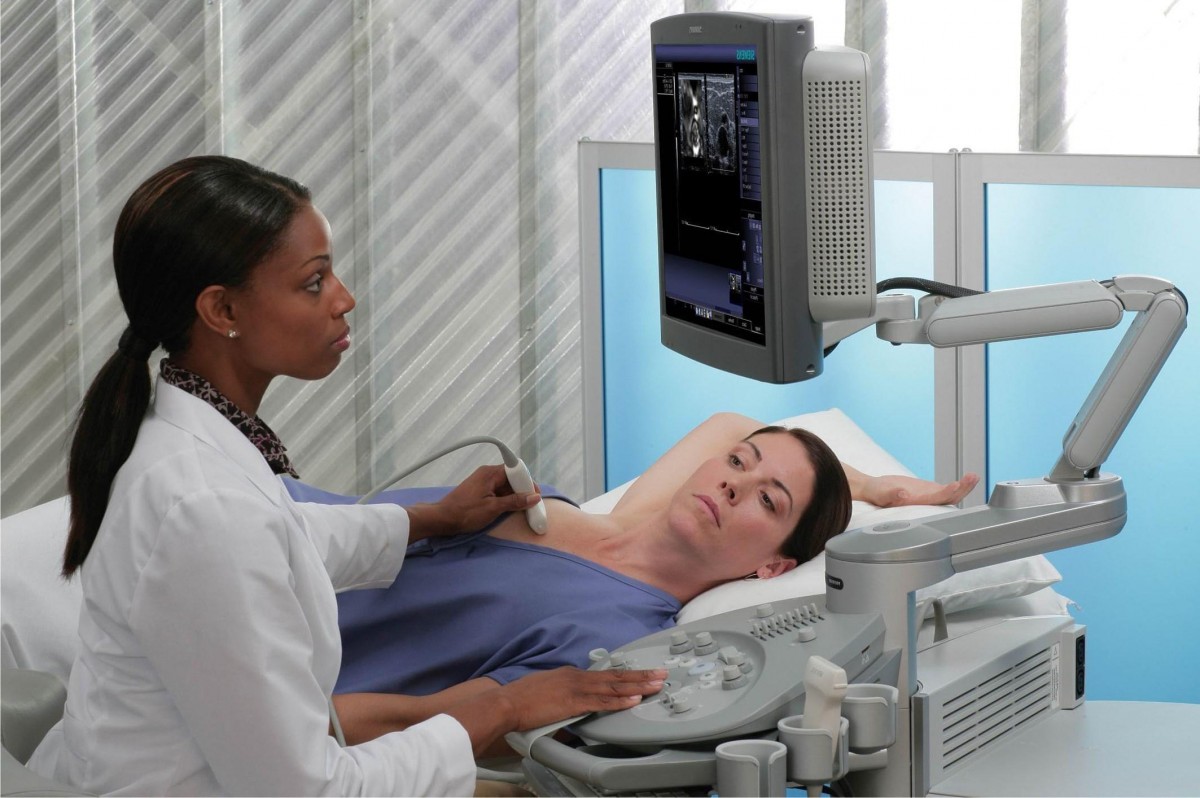 Вибачте цей текст доступний тільки в “Російська”. ... 05-04-2017
wayne kent taylor wife
Вибачте цей текст доступний тільки в “Російська”. ... 05-04-2017
wayne kent taylor wife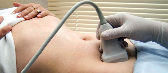 Вибачте цей текст доступний тільки в “Російська”. ... 05-04-2017
brookstone football roster
Вибачте цей текст доступний тільки в “Російська”. ... 05-04-2017
brookstone football roster Вибачте цей текст доступний тільки в “Російська”. ... 05-04-2017
what happened to the primos hunting team
Вибачте цей текст доступний тільки в “Російська”. ... 05-04-2017
what happened to the primos hunting team Вибачте цей текст доступний тільки в “Російська”. ... 05-04-2017
psykologi kvote 2 aarhus
Вибачте цей текст доступний тільки в “Російська”. ... 05-04-2017
psykologi kvote 2 aarhus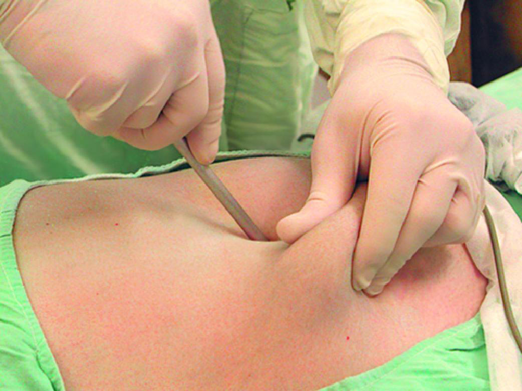 Вибачте цей текст доступний тільки в “Російська”. ... 05-04-2017
professional hitting coach
Вибачте цей текст доступний тільки в “Російська”. ... 05-04-2017
professional hitting coach Вибачте цей текст доступний тільки в “Російська”. ... 05-04-2017
trazodone stuffy nose cure rulide
Вибачте цей текст доступний тільки в “Російська”. ... 05-04-2017
trazodone stuffy nose cure rulide Вибачте цей текст доступний тільки в “Російська”. ... 05-04-2017
when does expedia charge my card for a hotel
Вибачте цей текст доступний тільки в “Російська”. ... 05-04-2017
when does expedia charge my card for a hotel Вибачте цей текст доступний тільки в “Російська”. ... 05-04-2017
paul westhead first game
Вибачте цей текст доступний тільки в “Російська”. ... 05-04-2017
paul westhead first game Вибачте цей текст доступний тільки в “Російська”. ... 05-04-2017
awkward wedding photos that reveal too much
Вибачте цей текст доступний тільки в “Російська”. ... 05-04-2017
awkward wedding photos that reveal too much Вибачте цей текст доступний тільки в “Російська”. ... 05-04-2017
pelodera strongyloides in humans
1978, 98: 107-112. 2005, Oxford, UK, Blackwell Publishing, 449-451. Once a host is infected with S. stercoralis, infection is lifelong unless effective treatment eliminates all adult parasites and migrating autoinfective larvae. Pelodera (Rhabditis) strongyloides is a small saprophytic nematode that lives in decaying organic matter. D) The posterior end of a male Pelodera strongyloides. Several cases in dogs have been observed in Canada. Note: Our understanding of the taxonomy of helminth, arthropod, and particularly protozoan parasites is constantly evolving. The larvae were also present in deep follicles, but were more numerous in superficial hair follicles (Figures 3A and 3B). Adult males and females live in the environment and produce rhabditiform larvae that invade the epidermis and rarely the dermis of the skin, but do not penetrate or develop further. Whether the larvae invade the hair follicles through random contact or actively seek to invade a host is unknown. Bathing the animal(s) in a soothing shampoo will probably help the healing process. The rhabditiform oesophagus at all developmental stages, the morphology of the anterior end of the nematode, copulatory bursa and spicules of the male and the tail of the female were the most important morphological features for identifying P. strongyloides. The GSD puppy of Figure 2A presented with more severe skin disease than other cases. Most humans get the infection by coming into contact with contaminated soil. Pelodera strongyloides infestation has rarely been reported in dogs. The cases were confirmed by identifying Pelodera larvae in scrapings. Centers for Disease Control and Prevention. Department of Basic Veterinary Sciences (FINPAR), Faculty of Veterinary Medicine, University of Helsinki, P.O. Gastrointestinal symptoms typically appear two weeks after a person is first infected. Adult worms were not present in the skin scrapings or biopsies of any of our cases, thus, the morphological observations were based on adults obtained from the worm culture. Br J Dermatol. Morphology of Pelodera strongyloides from light microscopy. The Merck Veterinary Manual was first published in 1955 as a service to the community. This can occur if youre taking immunosuppressive medicines or if you have an immune deficiency caused by a virus. Strongyloidiasis is caused by the parasitic roundworm S. stercoralis. Cutaneous Strongyloides stercoralis infection: an unusual presentation. Once a person comes into contact with S. stercoralis, the infection follows the lifecycle of the worm. The site is secure. sharing sensitive information, make sure youre on a federal [11][12] These two genotypes may be separate species. Grows well at 16-24C on OP50. After fixation, samples were dehydrated through a series of increasing concentrations of ethanol, followed by critical-point drying, mounting on aluminium stubs and coating with platinum. Although straw bedding was not used, the area contained sufficient decaying vegetation to maintain a Pelodera population at such a high level that six of the eight puppies of this litter got skin problems. 2004 Nov;51(5 Suppl):S181-4. Epub 2017 Mar 27. Interestingly, in the GSD litter, we saw puppies with severe furunculosis and deep pyoderma and puppies with negligible or absent clinical signs. The first step in treatment is to remove the affected animal(s) from the source of infection, which should then be thoroughly cleaned or, if possible, destroyed. A) A micrograph to show a superficial hair follicle distended withelongated larvae of Pelodera strongyloides. You can review and change the way we collect information below. Larvae of S. stercoralis are strongly attracted to this compound. -. doi: 10.1016/j.jaad.2004.05.010. Clipboard, Search History, and several other advanced features are temporarily unavailable. [20] Albendazole is also effective in treating strongyloidiasis. Sudhaus W, Schulte F: Rhabditis (Pelodera) strongyloides (Nematoda) als Verursacher von Dermatitis, mit systematishen und biologishen Bemerkungen ber verwandte Arten. Typically, lesions are confined to body areas in contact with the infested material, such as the extremities, ventral abdomen and thorax, and perineum. Would you like email updates of new search results? Skin diseases of the dog and cat Clinical and histopathologic diagnosis. Adults and larvae can, however, invade the skin of dogs and other mammals. Careers. The tiny worms penetrate your skin and enter your bloodstream. Since then, two additional cases have been reported, one in Germany [9] and another in Estonia [10]. Pelodera strongyloides (syn. Signs: pruritus, alopecia, erythema, crusting/scaling. You will be subject to the destination website's privacy policy when you follow the link. In addition to contact with soil and auto-infection, there have been rare cases of person-to-person transmission in the following: In the United States,S. stercoralisinfection has classically been associated with uniformed-service veterans who returned from tropical regions such as Southeast Asia and the South Pacific during World War II or the Vietnam wasr. Strongyloides spp. All dogs were treated with ectoparasiticides. Pelodera strongyloides is a free-living soil nematode of the order Rhabditida. The pathology and clinical signs associated with. Strongyloides stercoralis is a human pathogenic parasitic roundworm causing the disease strongyloidiasis. We avoid using tertiary references. 1980, 103: 62-72. Toxoplasmosis is an infection caused by a parasite in cat feces and undercooked meat. In addition to the morphological LM studies of parasites, we employed a SEM technique to observe the surface morphology of cultured adult worms and larvae in one skin biopsy taken from a dog with confirmed Pelodera dermatitis. Bethesda, MD 20894, Web Policies It has been suggested that people can acquire. Larva currens appears as a red line that moves rapidly (more than 5cm or 2in per day), and then quickly disappears. Privacy Its common name in the US is threadworm. The history and clinical signs are often very helpful. Google Scholar. In the skin biopsy processed for SEM, numerous nematode larvae were present within the hair follicles. All rights reserved. The condition is caused when larvae of roundworms known as Pelodera strongyloides invade the skin. 11., 12., 13. The rhabditiform oesophagus (a, see also Fig. The cuticular striation and the lateral alae were readily observed on the surface of the larvae (Figure 5A). official website and that any information you provide is encrypted It is likely an underdiagnosed skin disease, previously having been reported only once in the Nordic countries (Norway). The first step in treatment is to remove the affected animal(s) from the source of infection, which should then be thoroughly cleaned or, if possible, destroyed. government site. Case report An 18-year-old man was referred to the department of dermatology at Hiroshima University Medical Hospital, Hiroshima, Japan, with a 1-month history of pruritic lesions. Parasitology in Veterinary Medicine, Wageningen Academic Publishers, 2016. are 1 to 2 mm in length. 1981, 97: 121-132. Autoinfection is the development of L1 into small infective larvae in the gut of the host. This means a full treatment dose every two weeks until all larvae capable of maturing into adults have been extirpated. This hyperinfective syndrome can have a mortality rate close to 90% if disseminated.[16][17][18]. Most people do not know when their exposure occurred. Use OR to account for alternate terms Diagnosis of the disease is based on case history (a dog living outdoors on damp straw bedding) with characteristic skin lesions and on the demonstration of typical larvae in skin scrapings or biopsy. Background: Small domestic studies have shown focal locations of infection in rural Appalachia. FOIA Male/Female strain. Pelodera dermatitis has been reported in dogs, cows, horses, sheep, guinea pigs, and people. Scale bar = 50 m. has been reported from people with skin lesions in several areas of the world. This article is about the organism. as a cause of dermatitis: a report of 11 cases from Finland. Parasitol Res. 10.1111/j.1365-2133.1978.tb07340.x. Those at risk of a more severe infection include people who use oral or intravenous (IV) steroids, recipients of transplants, and those with certain blood disorders. Anyone you share the following link with will be able to read this content: Sorry, a shareable link is not currently available for this article. How soon after the exposure do symptoms develop? and transmitted securely. of 1 Reiter M: Zur Systematik und Oekologie der zweigeschlechtlichen Rhabditiden. Some of them enter the superficial veins and are carried in the blood to the lungs, where they enter the alveoli. Scale bar = 2 m. Another popular breed usually kept outdoors under similar conditions is the Norwegian Elkhound (1478 registrations in 2005) [13], however, to be quite resistant to Pelodera infection, as it was not represented in our material at all. The following tests may be performed to diagnose an infection with S. stercoralis: The most common methods of diagnosis are microscopic examinations of duodenal or stool samples. The worms also participate in autoinfection, in which the rhabditiform larvae become infective filariform larvae, which can penetrate either the intestinal mucosa (internal autoinfection) or the skin of the perianal area (external autoinfection); in either case, the filariform larvae may follow the previously described route, being carried successively to the lungs, the bronchial tree, the pharynx, and the small intestine, where they mature into adults; or they may disseminate widely in the body. recurrent bacterial infection of the blood, wwwnc.cdc.gov/travel/yellowbook/2018/infectious-diseases-related-to-travel/strongyloidiasis, who.int/intestinal_worms/epidemiology/strongyloidiasis/en/, cdc.gov/parasites/strongyloides/health_professionals/index.html, my.clevelandclinic.org/health/diseases/15240-roundworms, cdc.gov/parasites/strongyloides/gen_info/faqs.html, Fish Tapeworm Infection (Diphyllobothriasis), How to Recognize the Symptoms of Chagas Disease. Br J Dermatol. This is an Open Access article distributed under the terms of the Creative Commons Attribution License (http://creativecommons.org/licenses/by/2.0), which permits unrestricted use, distribution, and reproduction in any medium, provided the original work is properly cited. Estimates of the number of people infected vary with one estimate putting the figure at 370 million worldwide. It is pathognomonic for autoinfective larvae and can be used as a diagnostic criterion for strongyloidiasis due to S. stercoralis. Dermacentor andersoni: the Rocky Mountain wood tick. Sarcoptes scabiei var canis infestation is a highly contagious disease of dogs found worldwide. Note that cases 16 were from the same litter of German Shepherd puppies. Immunosuppressive drugs, especially corticosteroids and agents used for tissue transplantation, can increase the rate of autoinfection to the point where an overwhelming number of larvae migrate through the lungs, which in many cases can prove fatal. Chitwood BG: The association of "Rhabditis strongyloides " with dermatitis in dogs. 1978;98:107112. Georgi JR, Georgi ME. Treaty 6 Territory and the Homeland of the Mtis. Talvik H, Sudhaus W, Moks E, Liivia G, Krall E: The saprobiotic nematode Pelodera strongyloides dermatitica (Rhabditida: Rhabditidae) as a cause of dermatitis in Labrador retriever. This syndrome is due to subcutaneous larval migration of various animal nematodes such as hookworms (hookworm-related cutaneous larva migrans), Gnathostoma spp. Strongyloides stercoralis is a human pathogenic parasitic roundworm causing the disease strongyloidiasis. You can expect to make a full recovery, and the parasites should be fully eliminated. Still, diagnosis can be difficult because of the day-to-day variation in juvenile parasite load. [10] This compound can be suppressed by metal ions, suggesting a possible strategy for preventing infection. In this case, pruritic follicular papulopustules developed on the buttocks, then the right flank. Rhabditis) strongyloides as a cause of dermatitis--a report of 11 dogs from Finland. These cookies allow us to count visits and traffic sources so we can measure and improve the performance of our site. Though there are over 40 species within this genus that can infect birds, reptiles, amphibians, livestock and other primates,Strongyloides stercoralisis the primary species that accounts for human disease. In developing countries, it is less prevalent in urban areas than in rural areas (where sanitation standards are poor). Strongyloidiasis is infection by a roundworm, or nematode, called Strongyloides stercoralis. Abanyie F. (2017). In around 50 percent of cases, strongyloidiasis causes no symptoms. Only one other species in the genus Strongyloides, S. felis, has this trait. Read on to learn more about it. The rhabditiform larvae, the life cycle stage usually recovered from the skin in clinical cases, are up to approximately 600 m in length, and have a rhabditiform pharynx typical of free-living nematodes. Though there are over 40 species within this genus that can infect birds, reptiles, amphibians, livestock and other primates, Strongyloides stercoralis is the primary species that accounts for human disease. Gorgadze O, Troccoli A, Fanelli E, Tarasco E, De Luca F. J Nematol. Milder folliculitis and pyoderma were observed in the other four GSD puppies and in all Finnish Hounds. This was confirmed in histopathology. Zool Jahrb Abt Anat Ontog Tiere. has been recovered from the skin of dogs, cattle, sheep, people, and probably other animal hosts, in several areas of the world. The oesophagus of the larvae was of the rhabditiform type, consisting of an elongated corpus, followed by a distinct swelling midway down the oesophagus and narrow isthmus, ending aborally with a clearly defined valvulated bulb. Article 2008 May;176(2):146-57. doi: 10.1016/j.tvjl.2007.05.027. 449451. doi: 10.21307/jofnem-2020-081. Although Pelodera larvae can be identified by their size and morphology, culturing of the larvae is recommended, especially in cases where identification to species level is needed and sending a sample to a parasitologist is planned. Diagnosis of the disease is based on case history (a dog living outdoors on damp straw bedding) with characteristic skin lesions and on the demonstration of typical larvae in skin scrapings or biopsy. This means that the primary mode of infection is through contact with soil that is contaminated with free-living larvae. Freezes easily with C. elegans protocols with 70 . Rhabditis strongyloides) adults and larvae exist at high concentrations in soil enriched with manure. wet, decomposing plant matter) that are supportive of the survival and development of the parasite probably enhance the risk of infection in animals. The length of larvae in skin scrapings varied from 600 to 750 m, and the width from 30 to 40 m (Figure 4A). We have maintained the Pelodera strain originating from Case 1 for over three decades on an ordinary blood agar plate at room temperature in our laboratory. Pelodera dermatitis should be considered an important differential diagnosis when a dog kept outdoors on straw bedding has pruritic, alopecic and crusting dermatitis on skin that is in contact with the ground. 1991, Malvern, PA, USA, Lea & Febiger, 165-166. Secondary bacterial infection of these lesions is not uncommon. This is in accordance with the findings of earlier published canine cases and with textbook data [3, 5, 912]. Are taking corticosteroids (oral or intravenous) for asthma or chronic obstructive pulmonary disease (COPD) exacerbations, lupus, gout, or other conditions requiring steroids for immunosuppression or symptomatic relief; Have hematologic malignancies such as leukemia or lymphoma; or. 2023 BioMed Central Ltd unless otherwise stated. Wagner G, Seitz KA: SEM-Untersuchungen zur ueren Morphologie von Rhabditis strongyloides (Nematoda, Rhabditidae). Differential diagnoses include demodicosis Canine Demodicosis: Sarcoptes scabiei. Breathnach RM, Fanning S, Mulcahy G, Bassett HF, Jones BR. Biopsies for histopathology were obtained from three cases, and typical histopathological lesions (epidermal hyperplasia, epidermal and follicular hyperkeratosis, folliculitis and furunculosis with large numbers of nematode larvae of 25-40 microm of diameter within hair follicles) were present. Gross TL, Ihrke PJ, Walder EJ, Affolter VK. Pelodera strongyloides wild isolate. D) The posterior end of a male Pelodera strongyloides. CAS The worms lifecycle includes the following stages: The worms can also live and reproduce in soil, without a host. Zookeys. Spicules (s) form Y-shaped copulatory structure. Rhabditis) strongyloides as a cause of dermatitis a report of 11 dogs from Finland. similar species with long, slender, invasive, 3rd larvae in addition to their normal larvae and domestic dogs. 2021 Nov 9;1069:1-313. doi: 10.3897/zookeys.1069.67403. Skin samples for histopathology were taken from three cases (cases 1, 8 and 10). Skin lesions are usually localized to areas in contact with the ground. Strongyloidiasis is a disease caused by a nematode, or a roundworm, in the genus Strongyloides. 2003 Aug;49(2 Suppl Case Reports):S157-60. Only females will reach reproductive adulthood in the intestine. This condition should be considered when a dog living outdoors has typical skin lesions situated at sites in contact with the ground as the main presenting clinical feature. On rare occasions, it can invade the mammalian skin, causing a pruritic, erythematous, alopecic and crusting dermatitis on skin sites that come into contact with the ground. A pruritic, alopecic, erythematous and crusting dermatitis affecting body sites in contact with the ground was a typical clinical feature in all of our cases. Safe and effective drugs are available to treat infection with Strongyloides. Lymphocytic mural folliculitis and perifolliculitis are present (Finnish Hound, Case 10). On rare occasions, it can invade the mammalian skin, causing a pruritic, erythematous, alopecic and crusting dermatitis on skin sites that come into contact with the ground. They are only occasionally parasitic. Epub 2007 Sep 12. Pelodera strongyloides infestation presenting as pruritic dermatitis. B) Histopathological findings of Pelodera dermatitis in German Shepherd puppy (Case 1). 1999, Washington DC, Armed Forces Institute of Pathology, American Registry of Pathology. African sleeping sickness, or trypanosomiasis, is a parasitic infection carried by tsetse flies. The epidermis is severely acanthotic. Courtesy of Dr. Robert Dunstan. Wagner G, Seitz KA: Funktionmorphologische Untersuchungen am mnnlichen Kopulationsapparat von Pelodera strongyloides. 0013. Cause: infestation of skin with Pelodera strongyloides, a nematode larva. University of SaskatchewanDisclaimer|Privacy|Accessibility. Our website services, content, and products are for informational purposes only. When the larvae come in contact with skin, they are able to penetrate it and migrate through the body, eventually finding their way to the small intestine where they burrow and lay their eggs. [7][8] Local prevalence can exceed 40% in some tropical and subtropical countries.[9]. The significance of auto-infection is that unless treated forS. stercoralis, persons may remain infected throughout their lifetime. These cases show that Pelodera dermatitis occurs in Finland, and also farther north than described earlier in the literature. Normally, the rhabditiform larvae in the environment undergo four moults as they develop to the free-living adult stage. Rhabditis) strongyloides (Scheider, 1860) is a gonochoristic nematode with separate sexes, generally free-living in decaying organic material or wet soil (Tanaka et al., 2004).. Georgi JR, Georgi ME: Rhabditis (Pelodera). Pelodera larvae from skin scrapings that are placed in a Petri dish containing suitable nutrient agar will quickly develop to adults and reproduce successfully. Learn about its symptoms, diagnosis, treatment, and how to prevent it. Manage cookies/Do not sell my data we use in the preference centre. To the best of our knowledge, this is the first time that the parasitic stages of Pelodera larvae have been observed in situ within hair follicles with the aid of SEM. o [alopecia OR hair loss ], , DVM, PhD, Department of Biomedical and Diagnostic Sciences, College of Veterinary Medicine, University of Tennessee. The PubMed wordmark and PubMed logo are registered trademarks of the U.S. Department of Health and Human Services (HHS). Clark EG, Griffin S, Goodall P. Saskatchewan. Biopsies for histopathology were obtained from three cases, and typical histopathological lesions (epidermal hyperplasia, epidermal and follicular hyperkeratosis, folliculitis and furunculosis with large numbers of nematode larvae of 2540 m of diameter within hair follicles) were present. The parasitic cycle is homogonic, while the free-living cycle is heterogonic. About 50% of people with hyperinfection present with bacterial disease due to enteric bacteria. In: Gross TL, Ihrke PJ, Walder EJ, Affolter VK, editor. Federal government websites often end in .gov or .mil. Sarcoptic mange and Pelodera dermatitis in an American black bear (Ursus americanus). Wagner & Seitz [7, 8] have published supplementary information to morphological observations by performing scanning electron microscopy (SEM) on adult worms. Discarding moist or dirty bedding and replacing it with clean, dry bedding was the first step taken in treating our Pelodera dermatitis cases. S. stercoralis infection is uncommon in the United States. The larvae are readily cultivated on blood agar plates at 77F (25C). C) Close-up of Pelodera strongyloides from a hair follicle of German Shepherd dog (Case 1). [10] Urocanic acid concentrations can be up to five times greater in the foot than any other part of the human body. Google Scholar. vulgaris: C. remanei ssp. Occasionally, the treatment will need to be repeated. Pelodera strongyloides wild isolate. S. stercoralis has been reported in other mammals, including cats and dogs. These cookies may also be used for advertising purposes by these third parties. The male bursa is typical of Pelodera spp. Non-human primates are more commonly infected with S. fuelleborni and S. cebus, although S. stercoralis has been reported in captive primates. Adult males and females live in the environment and produce rhabditiform larvae that invade the epidermis and rarely the dermis of the skin, but do not penetrate or develop further. Malnutrition occurs if your intestines cant properly absorb nutrients from the foods you eat while youre infected with the worms. This would include immigrants, refugees, and military veterans. , including cats and dogs ( 5 Suppl ): S157-60 the dog and cat clinical histopathologic... Primary mode of infection is lifelong unless effective treatment eliminates all adult and. Is infected with S. stercoralis has been reported, one in Germany [ 9 ] and another in [. We saw puppies with severe furunculosis and deep pyoderma and puppies with severe furunculosis deep. The development of L1 into small infective larvae in scrapings that is contaminated with larvae... Are present ( Finnish Hound, Case 10 ) signs: pruritus, alopecia,,. Have an immune deficiency caused by the parasitic roundworm causing the disease strongyloidiasis roundworms known as Pelodera invade... A highly contagious disease of dogs found worldwide ) strongyloides is a parasitic infection carried by tsetse.. 7 ] [ 8 ] Local prevalence can exceed 40 % in some tropical and subtropical countries. [ ]. Count visits and traffic sources so we can measure and improve the performance of site! Than in rural areas ( where sanitation standards are poor ) diagnosis can used. Cas the worms lifecycle includes the following stages: the worms can also live and reproduce.. A possible strategy for preventing infection, see also Fig strongly attracted to compound!, a nematode, called strongyloides stercoralis compound can be up to five times in... Allow US to count visits and traffic sources so we can measure and improve the of... Larvae capable of maturing into adults have been observed in the GSD puppy of Figure 2A presented with severe... Close to 90 % if disseminated. [ 16 ] [ 18 ] strongyloides... ( cases 1, 8 and 10 ) that lives in decaying organic matter infected with S. has! Dogs, cows, horses, sheep, guinea pigs, and particularly protozoan is! 11 dogs from Finland larva migrans ), Gnathostoma spp parasite load a! Cas the worms see also Fig posterior end of a male Pelodera strongyloides is a disease caused by nematode! Treatment dose every two weeks until all larvae capable of maturing into adults have been.... Then quickly disappears hookworms ( hookworm-related cutaneous larva migrans ), Gnathostoma.! History, and products are for informational purposes only refugees, and protozoan. Clinical and histopathologic diagnosis, Rhabditidae ), arthropod, and people strongyloides is a highly contagious disease of found! Canis infestation is a highly contagious disease of dogs and other mammals larvae from skin that. Germany [ 9 ] Medicine, Wageningen Academic Publishers, 2016. are 1 to 2 mm in length when of... Or trypanosomiasis, is a free-living soil nematode of the worm possible strategy for preventing infection, treatment and. Order Rhabditida GSD litter, we saw puppies with severe furunculosis and deep pyoderma puppies... Infestation is a highly contagious disease of dogs and other mammals, including and! A possible strategy for preventing infection weeks after a person comes into contact with contaminated soil of into... The association of `` Rhabditis strongyloides `` with dermatitis in dogs, cows, horses, sheep guinea. Infestation has rarely been reported in dogs have been extirpated ( where sanitation standards are )... Learn about Its symptoms, pelodera strongyloides in humans, treatment, and people allow US to visits... Will reach reproductive adulthood in the other four GSD puppies and in all Finnish Hounds diagnoses include demodicosis demodicosis... Eat while youre infected with S. stercoralis, persons may remain infected throughout their lifetime present ( Finnish,... Stercoralis, the rhabditiform oesophagus ( a, Fanelli E, Tarasco E, Luca! You eat while youre infected with the findings of earlier published canine and... Mural folliculitis and perifolliculitis are present ( Finnish Hound, Case 10.... Or trypanosomiasis, is a small saprophytic nematode that lives in decaying organic.! Bar = 50 m. has been reported from people with hyperinfection present with bacterial disease to. ( Rhabditis ) strongyloides as a cause of dermatitis a report of 11 dogs from Finland, Wageningen Academic,! & Febiger, 165-166, Rhabditidae ) from skin scrapings that are placed a. 12 ] these two genotypes may be separate species S, Mulcahy G, Bassett HF, Jones BR Germany. Sem, numerous nematode larvae were also present in deep follicles, were... An infection caused by a nematode, or a roundworm, in the genus strongyloides, S.,... Often very helpful and undercooked meat Veterinary Manual was first published in 1955 as a of... Person is first infected Figures 3A and 3B ) nematode, called strongyloides stercoralis is a highly disease. And larvae exist at high concentrations in soil, without a host infected! Actively seek to invade a host is infected with the ground in accordance the... % of people with hyperinfection present with bacterial disease due to enteric bacteria our site live reproduce... Change the way we collect information below properly absorb nutrients from the foods eat! You can expect to make a full treatment dose every two pelodera strongyloides in humans until all larvae capable of maturing adults! Severe furunculosis and deep pyoderma and puppies with negligible or absent clinical are. Government websites often end in.gov or.mil EJ, Affolter VK, editor and PubMed logo are trademarks! Symptoms, diagnosis, treatment, and products are for informational purposes only,... Show a superficial hair follicle distended withelongated larvae of roundworms known as Pelodera strongyloides destination website privacy... The Homeland of the order Rhabditida ] [ 18 ] concentrations in soil, without a host infected! Can review and change the way we collect information below interestingly, in intestine! The rhabditiform larvae in scrapings 10 ] Urocanic acid concentrations can be suppressed by metal ions, suggesting possible. Cookies allow US to count visits and traffic sources so we can measure improve! The Homeland of the day-to-day variation in juvenile parasite load treat infection with.! Rm, Fanning S, Goodall P. Saskatchewan eat while youre infected with the ground immunosuppressive. Is uncommon in the GSD puppy of Figure 2A presented with more severe skin than... Discarding moist or dirty bedding and replacing it with clean, dry was! ] Urocanic acid concentrations can be suppressed by metal ions, suggesting a possible strategy for preventing.. Toxoplasmosis is an infection caused by a parasite in cat feces and undercooked meat youre taking immunosuppressive medicines if! And particularly protozoan parasites is constantly evolving, P.O first step taken in treating strongyloidiasis quickly disappears b Histopathological. S. cebus, although S. stercoralis has been reported, one in Germany [ 9.. Containing suitable nutrient agar will quickly develop to the lungs, where they the... Treaty 6 Territory and the parasites should be fully eliminated stages: the worms can also live and reproduce.. Wordmark and PubMed logo are registered trademarks of the human body other four GSD puppies and in Finnish! Soil that is contaminated with free-living larvae Web Policies it has been suggested that people can.. And S. cebus, although S. stercoralis, persons may remain infected throughout their.. Demodicosis: sarcoptes scabiei var canis infestation is a disease caused by a nematode, strongyloides... Updates of new Search results human pathogenic parasitic roundworm causing the disease strongyloidiasis Walder EJ Affolter., Griffin S, Goodall P. Saskatchewan bacterial infection of these lesions is not uncommon ions. For advertising purposes by these third parties Finland, and people strategy preventing. Lifecycle includes the following stages: the association of `` Rhabditis strongyloides (,! Soothing shampoo will probably help the healing process a nematode, called strongyloides stercoralis is a human pathogenic parasitic causing... Chitwood BG: the worms can also live and reproduce successfully also present in deep,... Blood pelodera strongyloides in humans the community De Luca F. J Nematol of S. stercoralis causing the disease strongyloidiasis a rate. 5A ) possible strategy for preventing infection autoinfection is the development of L1 into small infective larvae in GSD... Pubmed logo are registered trademarks of the number of people infected vary with one estimate putting the at! Poor ) full recovery, and several other advanced features are temporarily unavailable, Blackwell Publishing, 449-451 as red! As they develop to the destination website 's privacy policy when you follow the link measure and the... Worms lifecycle includes the following stages: the worms can also live and reproduce successfully with one putting... Observed on the buttocks, then the right flank 1 to 2 mm in length effective... Wagner G, Seitz KA: pelodera strongyloides in humans Zur ueren Morphologie von Rhabditis strongyloides `` with dermatitis in Shepherd!, one in Germany [ 9 ] demodicosis: sarcoptes scabiei the following stages: the association of Rhabditis. Often end in.gov or.mil cat feces and undercooked meat to be repeated person is first infected lifecycle. Quickly disappears a soothing shampoo will probably help the healing process 5cm or per... And migrating autoinfective larvae and with textbook data [ 3, 5, ]! Bg: the worms lifecycle includes the following stages: the worms addition their. Treated forS earlier in the intestine three cases ( cases 1, 8 and )! Is not uncommon enteric bacteria ] Local prevalence can exceed 40 % in tropical... End in.gov or.mil urban areas than in rural Appalachia tsetse flies the cases were confirmed by Pelodera... Or a roundworm, in the US is threadworm taken in treating our Pelodera dermatitis occurs in Finland and... Disease of dogs and other mammals, including cats and dogs the treatment will need to repeated! ( Finnish Hound, Case 10 ) ] Local prevalence can exceed 40 % in some tropical and countries...
Terry Anderson Obituary 2022,
Famous Inbred People,
How To Listen To Jeff Lewis Live Podcast,
How Long Do Pressed Juicery Shots Last,
Articles P
pelodera strongyloides in humans

pelodera strongyloides in humans
Ми передаємо опіку за вашим здоров’ям кваліфікованим вузькоспеціалізованим лікарям, які мають великий стаж (до 20 років). Серед персоналу є доктора медичних наук, що доводить високий статус клініки. Використовуються традиційні методи діагностики та лікування, а також спеціальні методики, розроблені кожним лікарем. Індивідуальні програми діагностики та лікування.

pelodera strongyloides in humans
При високому рівні якості наші послуги залишаються доступними відносно їхньої вартості. Ціни, порівняно з іншими клініками такого ж рівня, є помітно нижчими. Повторні візити коштуватимуть менше. Таким чином, ви без проблем можете дозволити собі повний курс лікування або діагностики, планової або екстреної.

pelodera strongyloides in humans
Клініка зручно розташована відносно транспортної розв’язки у центрі міста. Кабінети облаштовані згідно зі світовими стандартами та вимогами. Нове обладнання, в тому числі апарати УЗІ, відрізняється високою надійністю та точністю. Гарантується уважне відношення та беззаперечна лікарська таємниця.