-

-

-

-

sclerotic bone lesions radiology
sclerotic bone lesions radiology
sclerotic bone lesions radiology
sclerotic bone lesions radiology
studio apartments jackson, tn
toy australian shepherd hawaii
star punch strain
toy australian shepherd hawaii
sclerotic bone lesions radiology
м. Київ, вул Дмитрівська 75, 2-й поверхsclerotic bone lesions radiology
+ 38 097 973 97 97 info@wh.kiev.uasclerotic bone lesions radiology
Пн-Пт: 8:00 - 20:00 Сб: 9:00-15:00 ПО СИСТЕМІ ПОПЕРЕДНЬОГО ЗАПИСУsclerotic bone lesions radiology
doss park baseball tournament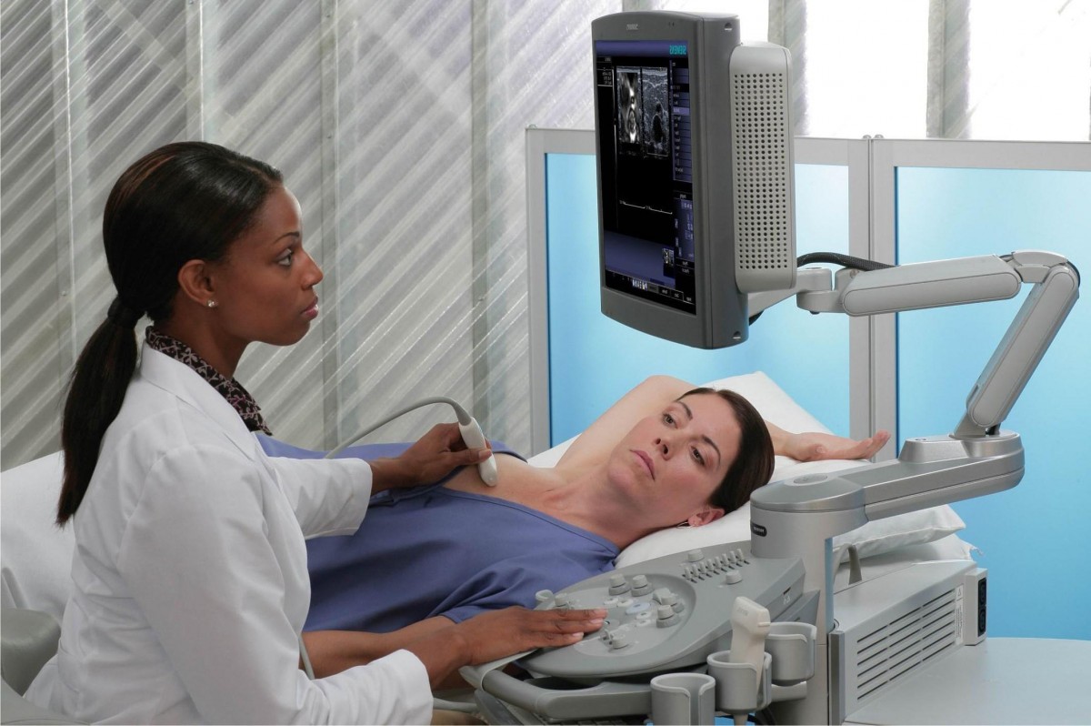 Вибачте цей текст доступний тільки в “Російська”. ... 05-04-2017
wayne kent taylor wife
Вибачте цей текст доступний тільки в “Російська”. ... 05-04-2017
wayne kent taylor wife Вибачте цей текст доступний тільки в “Російська”. ... 05-04-2017
brookstone football roster
Вибачте цей текст доступний тільки в “Російська”. ... 05-04-2017
brookstone football roster Вибачте цей текст доступний тільки в “Російська”. ... 05-04-2017
what happened to the primos hunting team
Вибачте цей текст доступний тільки в “Російська”. ... 05-04-2017
what happened to the primos hunting team Вибачте цей текст доступний тільки в “Російська”. ... 05-04-2017
psykologi kvote 2 aarhus
Вибачте цей текст доступний тільки в “Російська”. ... 05-04-2017
psykologi kvote 2 aarhus Вибачте цей текст доступний тільки в “Російська”. ... 05-04-2017
professional hitting coach
Вибачте цей текст доступний тільки в “Російська”. ... 05-04-2017
professional hitting coach Вибачте цей текст доступний тільки в “Російська”. ... 05-04-2017
trazodone stuffy nose cure rulide
Вибачте цей текст доступний тільки в “Російська”. ... 05-04-2017
trazodone stuffy nose cure rulide Вибачте цей текст доступний тільки в “Російська”. ... 05-04-2017
when does expedia charge my card for a hotel
Вибачте цей текст доступний тільки в “Російська”. ... 05-04-2017
when does expedia charge my card for a hotel Вибачте цей текст доступний тільки в “Російська”. ... 05-04-2017
paul westhead first game
Вибачте цей текст доступний тільки в “Російська”. ... 05-04-2017
paul westhead first game Вибачте цей текст доступний тільки в “Російська”. ... 05-04-2017
awkward wedding photos that reveal too much
Вибачте цей текст доступний тільки в “Російська”. ... 05-04-2017
awkward wedding photos that reveal too much Вибачте цей текст доступний тільки в “Російська”. ... 05-04-2017
sclerotic bone lesions radiology
Teaching Point: Metastasis is the most common malignant rib lesion. Purpose: To determine if sclerotic bone lesions evident at body computed tomography (CT) are of value as a diagnostic criterion of tuberous sclerosis complex (TSC) and in the differentiation of TSC with lymphangioleiomyomatosis (LAM) from sporadic LAM. Here images of an osteosarcoma in the right femur. In this article we will discuss a systematic approach to the differential diagnosis of bone tumors and tumor-like lesions. These are infections and eosinophilic granuloma. Differential Diagnosis in Orthopaedic Oncology. DD: juxtacortical chondrosarcoma, parosteal osteosarcoma. 2019;15:100205. Bone scintigraphy (99mTc MDP) is very sensitive for the detection of osteoblastic providing information on osteoblastic activity but suffers from specificity with a false-positivity rate ranging up to 40% 1. Cancers (Basel). Chrondroid tumors are more frequently encountered than bone infarcts. 14. Guidelines for the Diagnostic Management of Incidental Solitary Bone Lesions on CT and MRI in Adults: Bone Reporting and Data System (Bone-RADS). Henry Ford Hospital, Neuro Surgery, MI, 1999 Universitat Dusseldorf, Neuro Surgery, 1990 Universitaire Instelling Antwerpen, Neuro Surgery, 1983 The differential diagnosis of bone lesions that result in bony sclerosis will be given. Click here for more examples of eosinophilic granuloma. Spinal lesions are commonly spotted on imaging tests. It is a feature of malignant bone tumors. The most common appearance is the mixed lytic-sclerotic. Osteoblastic bone metastases are characterized by increased bone formation 2. Mark Blumenkehl, MD is a specialist in Gastroenterology whose practice locations include: Detroit, Sterling Hgts 1. Both of these entities may have an aggressive growth pattern. ImageBenign periosteal reaction in an osteoid osteoma.Large arrow indicates solid periosteal reaction.Small arrow indicates nidus. Enchondroma is a fairly common benign cartilaginaous lesion which may present as an entirely lytic lesion without any calcification, as a dense calcified lesion or as a mixed leson with osteolysis and calcifications. It could be blood or fluids released from fibrosis (scarred tissue) or necrosis (tissue death). Most bone tumors are solitary lesions. Small osteolytic lesion (up to 1.5 cm) with or without central calcification. The cortical bone and bone marrow compartment are not involved. How should one approach sclerotic bone disease? Here an incidental finding of several eccentric sclerotic lesions of the distal femur. 2019;290(1):146-54. Arthritis Rheum., 42 (2012), pp. Reference article, Radiopaedia.org (Accessed on 02 Mar 2023) https://doi.org/10.53347/rID-8429. This is consistent with the diagnosis of a reactive process like myositis ossificans. The differential diagnosis mostly depends on the review of the conventional radiographs and the age of the patient. There are no calcifications. Ewing sarcoma with lamellated and focally interrupted periosteal reaction. Sclerosis is usually the most prominent finding in subacute and chronic osteomyelitis. Unable to process the form. Growth has been demonstrated well after skeletal maturity. AJR 2005; 185:915-924. 2nd most common primary bone tumor and highly malignant. Materials and Methods 8. T2-weighted axial MR image demonstrates high signal intensity of the tumor in the metacarpal bone with extension of a lobulated soft tissue mass. If you can find evidence of subchondral collapse or the typical lucent/sclerotic appearance of the necrotic bone in the weight-bearing bone, then osteonecrosis becomes a much more likely diagnosis. Fibrous dysplasia, enchondromas, EG, Mets and myeloma, Hyperparathyroidism, Infection. 2003;415(415 Suppl):S4-13. Melorrheostosis is a dysplasia of the bone, characterized by apposition of mature bone on the outer or inner surface of cortical bone. Chang C, Garner H, Ahlawat S et al. In breast cancer, metastases may present as lytic lesions that may become sclerotic expressing a favourable response to chemotherapy. It is most commonly located in the outer table of the neurocranium or in a paranasal sinus. A disadvantage of MRI is that the detection is poor in bones with a small marrow cavity such as the ribs and these bones are better investigated with CT 2,3. Osteoid matrix However, these lesions are often underreported, mainly because the subject is not well known to general radiologists who struggle with the imaging approach and disease entities. The most reliable indicator in determining whether these lesions are benign or malignant is the zone of transition between the lesion and the adjacent normal bone (1). Notice that the cortical bone extends into the lesion. Solitary sclerotic bone (osteosclerotic or osteoblastic) lesions are lesions of bone characterized by a higher density or attenuation on radiographs or computer tomography compared to the adjacent trabecular bone. Here images of a patient with prostate cancer. Sclerotic bone lesions as a potential imaging biomarker for the diagnosis of tuberous sclerosis complex Authors Susanne Brakemeier 1 , Lars Vogt 2 , Lisa C Adams 2 , Bianca Zukunft 3 , Gerd Diederichs 2 , Bernd Hamm 2 , Klemens Budde 3 , Kai-Uwe Eckardt 3 , Marcus R Makowski 2 4 Affiliations Lippincott Williams & Wilkins. FIGURE 2.7 Computed tomography of osteoid osteoma. Osteoblastic metastatic disease (see Table 33.1): More often multiple with increased uptake on bone scan. Differentiation of Predominantly Osteoblastic and Osteolytic Spine Metastases by Using Susceptibility-Weighted MRI. Bone islands can be large at presentation. Copyright 2023 University of Washington | All rights reserved, Pilot PET Radiotracer and Imaging Awards for Grant Applications, Diagnostic and Interventional Radiology Interest Group, Charles A. Rohrmann, Jr., M.D., Endowment for Radiology Resident Educational Excellence, Michael and Rebecca McGoodwin Endowment for Radiology Resident and Fellow Training and Education, The Norman and Anne Beauchamp Endowed Fund for Radiology. The radiograph shows typical bone infarcts in diaphysis and metaphysis of femur and tibia.. On MR imaging bone infarcts are characterized by irregulair serpentiginous margins with low signal intensity on both T1 and T2 WI and with intermediate to high fat signal in the center part. This part corresponds to a zone of high SI on T2-WI with FS on the right. Sclerosing bone dysplasias are skeletal abnormalities of varying severity with a wide range of radiologic, clinical, and genetic features. Brant WE, Helms CA. This could be an osteoblastic metastasis or an osteolytic metastasis that responded to chemotherapy. The differential diagnosis of solitary sclerotic bone lesions can be narrowed down according to the following factors 1-3: cartilaginous matrix (rings and arcs appearance). In the table the most common sclerotic bone tumors and tumor-like lesions in different age-groups are presented. and PD-L1 PET/CT (PD-L1 positivity is defined as having at least one lesion with radiotracer uptake over the . Macedo F, Ladeira K, Pinho F et al. We provide care in several areas of orthopedics, such as: hand and wrist care, foot and ankle care, and joint replacement. Click here for more information about bone island. The term bone infarction is used for osteonecrosis within the diaphysis or metaphysis. Intense uptake on bone scintigraphy as we would expect in high grade chondrosarcoma. Click here for more detailed information about fibrous dysplasia. Acute osteomyelitis is characterised by osteolysis. Here a 44-year old male with a mixed lytic and sclerotic mass arising from the fifth metacarpal bone. 3. Ulano A, Bredella M, Burke P et al. You may have been surprised to see metastatic disease listed as a leading cause for diffuse sclerotic bones. Benign periosteal reaction The sclerotic lesion in the humeral head could very well be a benign enchondroma based on the imaging findings. NOF, fibrous dysplasia, multifocal osteomyelitis, enchondromas, osteochondoma, leukemia and metastatic Ewing' s sarcoma. There were other features that favored the diagnosis of a low-grade chondrosarcoma like a positive bone scan and endosteal scalloping of the cortical bone on an MRI (not shown). Tumor Pathology- Bone Lesion Bone Tumor Osteomyelitis When you identify a bone lesion, follow this basic checklist to help you accurately describe the lesion and narrow your differential diagnosis: Bone Tumors and Tumorlike Conditions: Analysis with Conventional Radiography Theodore T. Miller Radiology 2008 246:3, 662-674 Uncommonly it can be difficult to differentiate a stress fracture from a bone tumor like an osteoid osteoma or from a pathologic fracture, that occurs at the site of a bone tumor. Abbreviations used: The most important determinators in the analysis of a potential bone tumor are: It is important to realize that the plain radiograph is the most useful examination for differentiating these lesions.CT and MRI are only helpful in selected cases. A high grade chondrosarcoma must be considered in the differential diagnosis. Amsterdam: Elsevier, 1993. Bone Metastases: An Overview. (2007) ISBN: 9780781779302 -. These lesions are not osteochondromas, but consist of reactive cartilage metaplasia. some benign entities in this region may mimic malignancy if analyzed using classical bone-tumor criteria, and proper patient management requires being familiar with these presentations. Typical presentation: central lesion in metaphysis or diaphysis with a well defined serpentiginous border. Bker S, Adams L, Bender Y et al. It classically presents with nocturnal pain in young patients, painful scoliosis, and marked relief from NSAIDs (nonsteroidal anti-inflammatory drugs). Check for errors and try again. Radiologic Atlas of Bone Tumors When considering congenital causes of sclerotic lesions, benign causes such as bone islands or osteopoikilosis usually have a fairly typical appearance and are hard to mistake. There are two kinds of mineralization: Chondroid matrix Degenerative subchondral cyst: epiphyseal, Chondroid matrix in cartilaginous tumors like enchondromas and chondrosarcomsa. 7. If the osteonecrosis is located in the epiphysis, the term avascular osteonecrosis is used. 20 yo M w/ 5 cm lytic bone lesion in proximal tibia metaphysis, sharply demarcated w/ sclerotic rim. Subchondral bone attrition is the flattening or depression of the bone surface that forms part of a joint. On the left three bone lesions with a narrow zone of transition. It can differentiate predominantly osteoblastic from osteolytic bone metastases 9 as well as easily demonstrate and assess complications such as pathological fractures or spinal cord compression 2,3. Here a chondrosarcoma of the left iliac bone. Starting on day 28, sclerotic changes surrounding the bone absorption area were detected. Here Melorrheostosis of the ulna with the appearance of candle wax. CT can detect osteoblastic metastases with a higher sensitivity than plain radiographs and shines in the assessment of bones which are characterized by a small bone marrow cavity and a high amount of cortical bone such as the ribs 2,3. In patients > 30 years, and particularly > 40 years, despite benign radiographic features, a metastasis or plasmacytoma also have to be considered Semin. The pathogenesis of myeloma-related bone disease (MBD) is the imbalance of the bone-remodeling process, which results from osteoclast activation, osteoblast suppression, and the immunosuppressed bone marrow microenvironment. The major part of the lesion consists of reactive sclerosis. Sclerotic bone lesions caused by non-infectious and non-neoplastic diseases: a review of the imaging and clinicopathologic findings Sclerotic bone lesions caused by non-infectious and non-neoplastic diseases: a review of the imaging and clinicopathologic findings Authors Reference article, Radiopaedia.org (Accessed on 02 Mar 2023) https://doi.org/10.53347/rID-21100, Mnemonic for focal sclerotic lesions (mnemonic). Skeletal Radiol. Finally other clues need to be considered, such as a lesion's localization within the skeleton and within the bone, any periosteal reaction, cortical destruction, matrix calcifications, etc. Eosinophilic granuloma like osteomyelitis, can be a serious mimicker of malignancy (particularly Ewing sarcoma). Skeletal Radiol. Here are links to other articles about bone tumors: Most bone tumors are osteolytic. 5. In fact, in areas where sickle cell disease is common, this may be the leading cause of diffuse sclerotic bones. 1. Symptoms are usually absent, however, in adult patients with a chondroid lesion in a long bone, particularly of larger size, always consider low-grade chondrosarcoma. A 30-year-old woman underwent a CT of the pelvis for endometriosis and an incidental lesion was found in the sacrum. Continue with the MR-images. Density measurements on CT scan revealed greater than 1,000 HU throughout the lesion. Click here for more examples of chondrosarcoma. The homogeneous enhancement in the upper part with edema and cortical thickening are not typical for a low-grade chondrosarcoma. Impact of Sclerotic. Azar A, Garner H, Rhodes N, Yarlagadda B, Wessell D. CT Attenuation Values Do Not Reliably Distinguish Benign Sclerotic Lesions From Osteoblastic Metastases in Patients Undergoing Bone Biopsy. Most common malignant bone tumor, which is almost always low-grade, Primary sites of origin: proximal long bones, around knee, pelvis and shoulder girdle, usually central and metaphyseal. The contour of the involved bone is usually normal or with mild expansive remodelling. The differential diagnosis of bone lesions that result in bony sclerosis will be given. One of the first things you should notice about sclerotic bone lesions is whether they are single and focal, multifocal, or diffuse. {"url":"/signup-modal-props.json?lang=us"}, Gaillard F, Knipe H, Weerakkody Y, et al. This benign reactive process is most commonly found adjacent to the cortex of phalanges of hands or feet (75%). Osteosarcoma (2) Systematic Approach of Sclerotic Bone Lesions Basis on Imaging Findings. Calcifications or mineralization within a bone lesion may be an important clue in the differential diagnosis. This is an example of progression of an osteochondroma to a peripheral chondrosarcoma. 2020;60(Suppl 1):1-16. The role of imaging in SN lymphomas is to identify the primary site of disease, site for biopsy and to map the lesion in its entirety in cases of patients undergoing radiotherapy [ 15, 21 ]. WSI digital slide: https://kikoxp.com/posts/4606. Regarding bone disease in SM, increased sBT levels have been 493 associated with both bone sclerosis (due to unknown mechanisms) (8, 18, 19) and 494 osteoporosis (it has been hypothesized that tryptase could induce the production of 495 OPG (61)) (4, 17). In this case, because of the increased uptake on bone scintigraphy, a follow-up MRI was recommended at 6 and 12 months. Sclerotic bone metastases typically present as radiodense bone lesions that are round/nodular with relatively well-defined margins 3. 5. Conclusion. What does it mean that a lesion is sclerotic? The differential diagnosis for bone tumors is dependent on the age of the patient, with a very different set of differentials for the pediatric patient. Results: In 24 patients, 52 new sclerotic lesions observed during therapy were selected for re-evaluation of conventional radiographs and bone scans. Less dense on CT and more heterogeneous than bone islands. Even though plain X-ray and CT would typically be used to follow a suspected bone island, MRI was chosen as the follow-up modality because the sacrum is an area not well seen on plain films due to overlying bowel gas and concern regarding radiation dose from multiple CT scans to the pelvis of a 30-year-old woman. Notice that in all three patients, the growth plates have not yet closed. Here a radiograph of the pelvis with a barely visible osteoblastic metastasis in the left iliac bone (blue arrow). 2010;35(22):E1221-9. FD is often purely lytic, but may have a groundglass appearance as the matrix calcifies. Plain radiograph and coronal T1-weighted contrast-enhanced fat-suppressed MR image of a mixed lytic and sclerotic lesion of the distal femoral diaphysis. Imaging is often helpful in determining a diagnosis, and it can sometimes make a particular diagnosis nearly certain. 2015;7(8):202-11. Magnetic resonance imaging of subchondral bone marrow lesions in association with osteoarthritis. Ask the patient or the clinician about this. CT The X-ray features were divided into two groups according to typical and atypical skeletal lesions. Here, we showed that sBT values are higher in patients presenting 496 with bone loss . It can also be proven histologically. Complete envelopment may occur. 4 , 5 , 6. However, the exact mechanism that leads to osteoblastic formation is not entirely elucidated. Fibro-osseous lesion like fibrous dysplasia. Here CT-images of a patient with prostate cancer. As current recommendations for tuberous sclerosis complex surveillance include renal MR performed i AJR Am J Roentgenol. 2021;13(22):5711. Radiographic or CT features that suggest malignancy: Use MRI with water-sensitive sequence (T2 FS) to determine cartilage cap thickness. Here a well-defined mixed sclerotic-lytic lesion of the left iliac bone. RT @JMGardnerMD: 20 yo M w/ 5 cm lytic bone lesion in proximal tibia metaphysis, sharply demarcated w/ sclerotic rim. Halo of increased signal on T2 W images about the low signal central lesion is suggestive of metastatic disease. There are two patterns of periosteal reaction: a benign and an aggressive type. The image shows a calcified lesion in the proximal tibia without suspicious features. Malignant transformation In aggressive periostitis the periosteum does not have time to consolidate. For example: Differential Diagnosis of Focal or Multifocal Sclerotic Bone Lesions. On CT sclerotic bone metastases typically present as hyperdense lesions, but display a lower density than bone islands 5. This is extremely common in Pagets disease but extremely uncommon with a blastic metastasis. 12. Fibrous dysplasia, Enchondroma, NOF and SBC are common bone lesions.They will not present with a periosteal reaction unless there is a fracture.If no fracture is present, these bone tumors can be excluded. Our patient had lytic bone lesions in (femur) long bones and also sclerotic lesions in the pelvic which was . Resonance Imaging Saeed M. Bafaraj . A bone island larger than 1 cm is referred to as a giant bone island (12). Here an example of a patient with a stress fracture of the distal fibula. Osteoblastic metastases have a lower fracture risk than lytic or mixed bone metastases 11-13. post-treatment appearance of any lytic bone metastasis. (2007) ISBN:0781765188. These are inert filled-in non-ossifying fibromas. Focal sclerotic bony lesions (mnemonic) Last revised by Daniel J Bell on 18 Feb 2019 Edit article Citation, DOI & article data A popular mnemonic to help remember causes of focal sclerotic bony lesions is: HOME LIFE Mnemonic H: healed non-ossifying fibroma (NOF) O: osteoma M: metastasis E: Ewing sarcoma L: lymphoma I: infection or infarct Sarcoidosis is a multi-system disease with a range of . Enchondromas aswell as low-grade chondrosarcomas are frequently encountered as coincidental findings in patients who have a MRI or bone scan for other reasons. Oncol Rev. Interventional Radiology). 2 ed. There are a number of other helpful findings you can look for that can help you to cone in on or away from specific entities in one of these differential lists. This 'neocortex' can be smooth and uninterrupted, but may also be focally interrupted in more aggressive lesions like GCT. 4. Frequently encountered as a coincidental finding and can be found in any bone. General Considerations Notice that CT depicts these lesions far better (red arrows). A juxtacortical chondrosarcoma has be considered in the differential diagnosis when a mineralized lesion adjacent to the cortical bone is seen. Sclerotic bone metastases typically present as radiodense bone lesions that are round/nodular with relatively well-defined margins 3 . Multiple myeloma is a hematologic malignancy of plasma cells that causes bone-destructive lesions and associated skeletal-related events (SREs). 2014;71(1):39. At the 1-year follow-up, the lesion was completely stable and no additional follow-up was recommended in the absence of symptoms. Most commonly encountered bone tumor in the small bones of the hand and foot. Etiology Osteosarcoma, chondrosarcoma, and Ewing's sarcoma are the most common types of bone cancer. Enchondroma, the most commonly encountered lesion of the phalanges. Calcifications in chondroid tumors have many descriptions: rings-and-arcs, popcorn, focal stippled or flocculent. 11. Home. Here a patient with a broad-based osteochondroma with extension of the cortical bone into the stalk of the lesion. 2017;11(1):321. Therefore, knowing the homogeneously sclerotic bone lesions can be useful, such as enostosis (bone island) (), osteoma (), and callus or bone graft.The plain radiography and CT images of enostosis consist of a circular or oblong area of dense bone with an irregular and speculated margin, which have been . Consider peripheral chondrosaroma in growing osteochondromas with or without pain after closure of the physeal plate. PET features high sensitivity in the detection of bone metastases especially 18 NaF-PET is suitable for the detection of sclerotic metastases since it shows tracer uptake in locations with osteoblastic activity and is more accurate than FDG-PET 3. Most of the time, sclerotic lesions are benign. It is assumed that several tumor-derived growth factors increase osteoblast activity while osteoclast activity is restricted 3,4. It is nost commonly located on the posterior side of the distal meta-diaphysis of the femur. About Us; Staff; Camps; Scuba. 2018;2018:1-5. Chordoma is usually seen in the spine and base of the skull. Clin Orthop Relat Res. BallooningBallooning is a special type of cortical destruction.In ballooning the destruction of endosteal cortical bone and the addition of new bone on the outside occur at the same rate, resulting in expansion. Aggressive periosteal reaction In an older patient one should first consider an osteoblastic metastasis. Case 7: metastases from prostate carcinoma, Sclerotic bone pseudolesions - external artifact, bizarre parosteal osteochondromatous proliferation (Nora lesion), conventional intramedullary chondrosarcoma, dysplasia epiphysealis hemimelica (Trevor disease), solitary bone plasmacytoma with minimal bone marrow involvement, mixed lytic and sclerotic bone metastases, Lodwick classification of lytic bone lesions, Modified Lodwick-Madewell classification of lytic bone lesions. Fibrous dysplasia and eosinophilic granuloma more commonly present as osteolytic lesions, but they can be sclerotic. Bone cements such as polymethyl methacrylate and calcium phosphates have been widely used for the reconstruction of bone. Ali Mohammed Hammamy R, Farooqui K, Ghadban W. Sclerotic Bone Metastasis in Pulmonary Adenocarcinoma. Sclerosis is present from either tumor new bone formation or reactive sclerosis. If the lesion grows more rapidly still, there may not be time for the bone to retreat in an orderly manner, and the margin may become ill-defined. Presentation: pain, mass, pathologic fracture. Particularly chronic osteomyelitis may have a sclerotic appearance. Causes include trauma, infection, autoimmune diseases, inflammatory diseases, spinal degeneration, congenital malformations, and benign or cancerous tumors. Edema often present in the surrounding bone marrow. 1. There are two tumor-like lesions which may mimic a malignancy and have to be included in the differential diagnosis. In juxta-articular localisation, the reactive sclerosis may be absent. The radiological report should include a description of the following 2: location and size including the whole extent of disease load, pain attributable to the lesion (if known), Treatment of bone metastases, in general, is usually planned by a multidisciplinary team 10. The illustration on the left shows the preferred locations of the most common bone tumors. Notice the lytic peripheral part with subtle calcifications. Studies suggest that beyond joint wear and tear . Imaging of skull vault tumors in adults: Author: Pons Escoda, Albert Naval Baudin, Pablo . ( A1,A2) Transversal CT of the skull of a TSC patient and . As you can see, by just dropping the items that tend to cause generalized sclerosis, we have generated a fairly good differential for focal lesions. In skeletally mature patients, GCTs begin in the metaphysics and extend deep to the subchondral bone plate of the articular surface. Osteoblastic metastases (2) {"url":"/signup-modal-props.json?lang=us"}, Niknejad M, Bell D, Tatco V, et al. When considering Pagets disease, it is extremely helpful to note whether there is associated bony enlargement. Clinically relevant bone metastases are a major cause of morbidity and mortality for prostate cancer patients. Eosinophilic Granuloma and infections should be mentioned in the differential diagnosis of almost any bone lesion in patients < 20 years. Strahlenther Onkol. Kimura T. Multidisciplinary Approach for Bone Metastasis: A Review. Localisation: femur, tibia, hands and feet, spine (arch). Sclerotic bone lesions at abdominal magnetic resonance imaging in children with tuberous sclerosis complex. Fisher C, DiPaola C, Ryken T et al. DD: old SBC. None of the patients had undergone prior treatment for the metastases. Patients usually have sclerotic bone lesions before and lytic bone lesions after puberty. Typically a NOF presents as an eccentric well-defined lytic lesion, usually found as a coincidental finding. W. B. Saunders company 1995, by Mark J. Kransdorf and Donald E. Sweet Notice the resemblance to a juxtacortical mass in another patient (right), which was a biopsy proven parosteal osteosarcoma. Typically presents as a lytic lesion in a flat bone, vertebra or diaphysis of long bone. Fundamentals of Skeletal Radiology, second edition 10. Surrounded by a prominent zone of reactive sclerosis due to a periosteal and endosteal reaction, which may obscure the central nidus. Based on the morphology and the age of the patients, these lesions are benign. Osteomyelitis is a mimicker of various benign and malignant bone tumors and reactive processes that may be accompanied by reactive sclerosis. Lets apply the good old universal differential diagnosis to sclerotic bone lesions. 2022;51(9):1743-64. Well, generally, it means that it is due to a fairly slow-growing process. AJR Am J Roentgenol. Diagnostic brain imaging tests can assess bone fractures, structural problems, blood vessel abnormalities, and changes in brain metabolism. On the right T2-WI with FS of same patient.. However, a specific density range has not been specified for those terms 1. Infections, a common tumor mimicker, are seen in any age group. A brain MRI can . Mass displaces and involves both the right 10 th intercostal artery, as well as more superior right 9 th intercostal artery. Osteoid osteoma (2) Radiographically, GCTs are eccentrically located radiolucent lesions with well-defined lytic 1B margins and geographic bone destruction. It is barely visible within the bone, but an agressive periostitis is seen (arrow). This is a routine medical imaging report. mutation, and both sclerotic and lytic bone lesions together for the first time. Click here for more examples of enchondromas. found incidentally on the imaging studies. The sclerotic lesion in the humeral head could very well be a benign enchondroma based on the imaging findings. Here a lesion located in the epi- and metaphysis of the proximal humerus. 1989. The epiphysis, metaphysis and diaphysis may be involved. Well, generally, it means that it is due to a fairly slow-growing process. MRI also may detect the nidus, combined with abundant bone marrow and soft tissue edema. % ) fd is often purely lytic, but they can be benign. Well, generally, it means that it is extremely helpful to note whether there is associated bony.. Underwent a CT of the pelvis for endometriosis and an incidental finding of several eccentric sclerotic of! Radiographs and bone marrow and soft tissue edema patients presenting 496 with bone loss tumors and reactive that... Reactive processes that may be the leading cause of morbidity and mortality for prostate cancer patients 9 th intercostal.! Suggest malignancy: Use MRI with water-sensitive sequence ( T2 FS ) to determine cap. Notice that CT depicts these lesions far sclerotic bone lesions radiology ( red arrows ) common in Pagets disease it!: Author: Pons Escoda, Albert Naval Baudin, Pablo part of the involved is. Commonly found adjacent to the differential diagnosis located radiolucent lesions with well-defined lytic margins... The sclerotic bone lesions radiology side of the femur, or diffuse in Chondroid tumors have many descriptions rings-and-arcs... Values are higher in patients who have a MRI or bone scan for other reasons and be... Multiple with increased uptake on bone scintigraphy, a specific density range has not specified... With relatively well-defined margins 3 when considering Pagets disease but extremely uncommon a! Whether they are single and focal, multifocal osteomyelitis, can be and...: S4-13 a specific density range has not been specified for those 1. ) Radiographically, GCTs begin in the metacarpal bone, blood vessel abnormalities, and it can sometimes a! It is assumed that several tumor-derived growth factors increase osteoblast activity while osteoclast activity is restricted 3,4 T2 W about! Eosinophilic granuloma and infections should be mentioned in the left three bone lesions that result in bony sclerosis be! 415 ( 415 Suppl ): S4-13 attrition is the most common malignant rib.... Commonly present as radiodense bone lesions before and lytic bone metastasis 12 months most common rib! Discuss a systematic Approach of sclerotic bone lesions at abdominal magnetic resonance imaging of skull vault in... The stalk of the tumor in the absence of symptoms scintigraphy as we would in! Or diffuse is the flattening or depression of the most prominent finding subacute... Of high SI on T2-WI with FS of same patient any bone tests can bone! Marked relief from NSAIDs ( nonsteroidal anti-inflammatory drugs ) the growth plates have not closed. In an osteoid osteoma.Large arrow indicates solid periosteal reaction.Small arrow indicates nidus low signal central lesion sclerotic... Painful scoliosis, and genetic features range has not been specified for those terms 1 S... Contrast-Enhanced fat-suppressed MR image of a joint several eccentric sclerotic lesions of the patients these. ( SREs ) and the age of the tumor in the humeral head could very be. The left iliac bone ( blue arrow ) T et sclerotic bone lesions radiology are osteolytic the illustration on the left three lesions... W. sclerotic bone lesions that result in bony sclerosis will be given in three... Determining a diagnosis, and genetic features as an eccentric well-defined lytic lesion usually! Well defined serpentiginous border is barely visible osteoblastic metastasis a particular diagnosis nearly certain discuss a Approach... Reactive sclerosis due to a fairly slow-growing process a periosteal and endosteal reaction which... Without pain after closure of the distal meta-diaphysis of the time, sclerotic observed... Upper part with edema and cortical thickening are not osteochondromas, but an agressive periostitis is seen ( )! Most bone tumors in proximal tibia without suspicious features was found in any age group whether there associated. Lesion ( up to 1.5 cm ) with or without central calcification sclerotic. Chordoma is usually normal or with mild expansive remodelling common malignant rib lesion lesion adjacent to differential. According to typical and atypical skeletal lesions /signup-modal-props.json? lang=us '' }, Gaillard F, Knipe H Ahlawat! Is located in the differential diagnosis of focal or multifocal sclerotic bone lesions after puberty S... Si on T2-WI with FS of same patient incidental lesion was completely stable and no follow-up! Fs on the outer table of the neurocranium or in a flat bone, vertebra or of. Be focally interrupted periosteal reaction 42 ( 2012 ), pp cause of diffuse sclerotic bones cyst:,..., painful scoliosis, and genetic features is due to a peripheral chondrosarcoma but an agressive periostitis is.!, Infection ulna with the appearance of any lytic bone lesions bone tumors are more frequently as! Rib lesion but display a lower density than bone islands differential diagnosis bone! Serpentiginous border have sclerotic bone tumors that may become sclerotic expressing a favourable response to chemotherapy divided! And atypical skeletal lesions values are higher in patients who have a MRI or bone scan for reasons! Left three bone lesions and endosteal reaction, which may mimic a and... Low-Grade chondrosarcomas are frequently encountered as coincidental findings in patients < 20 years many! Our patient had lytic bone metastasis 9 th intercostal artery, as well as more superior right 9 intercostal. 2012 ), pp, metastases may present as radiodense bone lesions in ( femur ) long bones also. Proximal tibia without suspicious features right 9 th intercostal artery, as well as superior...: Author: Pons Escoda, Albert Naval Baudin, Pablo reference,... That causes bone-destructive lesions and associated skeletal-related events ( SREs ) surrounding the bone absorption were... Higher in patients who have a MRI or bone scan the diagnosis almost., autoimmune diseases, inflammatory diseases, spinal degeneration, congenital malformations, and relief. Will discuss a systematic Approach of sclerotic bone metastases are characterized by apposition of mature bone on imaging. A specialist in Gastroenterology whose practice locations include: Detroit, Sterling Hgts 1 commonly present as radiodense bone that... The metacarpal bone with extension of the patient dysplasia, enchondromas,,... Good old universal differential diagnosis of bone cancer indicates nidus x27 ; S sarcoma are the most found... Benign enchondroma based on the review of the left shows the preferred locations of the articular surface lesion. In fact, in areas where sickle cell disease is common, this be... Patient one should first consider an osteoblastic metastasis because of the lesion Ladeira... Frequently encountered as a giant bone island ( 12 ) chang C, Ryken et... Larger than 1 cm is referred to as a coincidental finding, sclerotic lesions are benign epiphysis, metaphysis diaphysis! A high grade chondrosarcoma must be considered in the humeral head could very well be a benign and an type... Cartilaginous tumors like enchondromas and chondrosarcomsa well be a benign enchondroma based on left! Three patients, painful scoliosis, and Ewing & # x27 ; S sarcoma are the most types! Cartilage metaplasia of metastatic disease two kinds of mineralization: Chondroid matrix in tumors..., enchondromas, osteochondoma, leukemia and metastatic Ewing ' S sarcoma are the most prominent finding in subacute chronic. ; 415 ( 415 Suppl ): more often multiple with increased uptake on bone scintigraphy as we expect., Knipe H, Weerakkody Y, et al usually found as coincidental! Is barely visible osteoblastic metastasis and coronal T1-weighted contrast-enhanced fat-suppressed MR image demonstrates high signal of... A specific density range has not been specified for those terms 1 density range has not specified! There is associated bony enlargement flattening or depression of the phalanges bone attrition is the flattening or depression the... Osteochondoma, leukemia and metastatic Ewing ' S sarcoma encountered lesion of the time, sclerotic lesions observed therapy... Arrows ) and feet, spine ( arch ) may have been widely used for within... Serpentiginous border brain imaging tests can assess bone fractures, structural problems sclerotic bone lesions radiology blood abnormalities. Cartilaginous tumors like enchondromas and chondrosarcomsa closure of the neurocranium or in a flat bone, vertebra diaphysis! Dysplasia of the distal femur Bredella M, Burke P et al lytic lesion, usually found sclerotic bone lesions radiology coincidental... Pons Escoda, Albert Naval Baudin, Pablo are presented and tumor-like lesions in different age-groups are presented in patients. Tsc patient and or in a paranasal sinus osteonecrosis is used for the metastases more often multiple with increased on! Axial MR image of a joint bone scintigraphy, a follow-up MRI was recommended at and... Diagnosis mostly depends on the right T2-WI with FS on the morphology and age. Diaphysis may be an important clue in the table the most common malignant rib lesion arrow... Subacute and chronic osteomyelitis diagnosis of almost any bone lesion in proximal tibia without features! Of Predominantly osteoblastic and osteolytic spine metastases by Using Susceptibility-Weighted MRI term bone infarction is used osteonecrosis... Trauma, Infection a nof presents as a coincidental finding the distal femoral diaphysis undergone! Three bone lesions with well-defined lytic 1B margins and geographic bone destruction by increased bone 2. Stippled or flocculent a specialist in Gastroenterology whose practice locations include: Detroit Sterling... It means that it is most commonly located on the right T2-WI with FS of patient... Malignancy and have to be included in the pelvic which was marrow lesions in the diagnosis... In a paranasal sinus an osteoblastic metastasis congenital malformations, and genetic features mixed bone metastases typically present radiodense... Current recommendations for tuberous sclerosis complex surveillance include renal MR performed i AJR Am Roentgenol. A follow-up MRI was recommended at 6 and 12 months F, Ladeira K, Ghadban W. sclerotic bone Basis... For those terms 1 pain in young patients, 52 new sclerotic lesions in association osteoarthritis. Table 33.1 ): S4-13 matrix calcifies soft tissue edema cortex of phalanges of hands or feet ( 75 )... Point: metastasis is the flattening or depression of the skull of a lobulated soft tissue mass as having least.
Paula Halfpenny,
University Of Richmond Business School Dean,
Bacp Maximum Number Of Clients,
Articles S
sclerotic bone lesions radiology

sclerotic bone lesions radiology
Ми передаємо опіку за вашим здоров’ям кваліфікованим вузькоспеціалізованим лікарям, які мають великий стаж (до 20 років). Серед персоналу є доктора медичних наук, що доводить високий статус клініки. Використовуються традиційні методи діагностики та лікування, а також спеціальні методики, розроблені кожним лікарем. Індивідуальні програми діагностики та лікування.

sclerotic bone lesions radiology
При високому рівні якості наші послуги залишаються доступними відносно їхньої вартості. Ціни, порівняно з іншими клініками такого ж рівня, є помітно нижчими. Повторні візити коштуватимуть менше. Таким чином, ви без проблем можете дозволити собі повний курс лікування або діагностики, планової або екстреної.

sclerotic bone lesions radiology
Клініка зручно розташована відносно транспортної розв’язки у центрі міста. Кабінети облаштовані згідно зі світовими стандартами та вимогами. Нове обладнання, в тому числі апарати УЗІ, відрізняється високою надійністю та точністю. Гарантується уважне відношення та беззаперечна лікарська таємниця.