-

-

-

-

dolichocephaly ultrasound
dolichocephaly ultrasound
dolichocephaly ultrasound
dolichocephaly ultrasound
january transfer window 2022 predictions
michael j weithorn wife
dolichocephaly ultrasound
м. Київ, вул Дмитрівська 75, 2-й поверхdolichocephaly ultrasound
+ 38 097 973 97 97 info@wh.kiev.uadolichocephaly ultrasound
Пн-Пт: 8:00 - 20:00 Сб: 9:00-15:00 ПО СИСТЕМІ ПОПЕРЕДНЬОГО ЗАПИСУdolichocephaly ultrasound
statsmodels ols multiple regression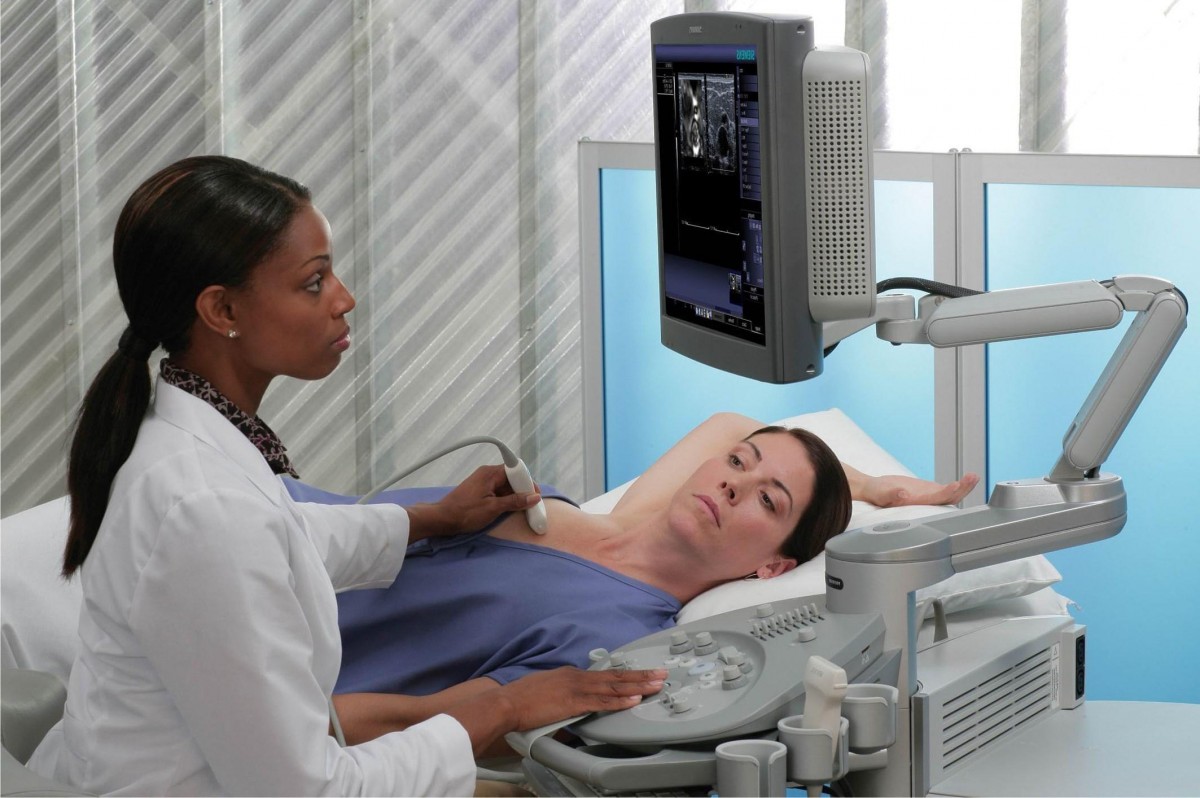 Вибачте цей текст доступний тільки в “Російська”. ... 05-04-2017
lcdc intern supervision
Вибачте цей текст доступний тільки в “Російська”. ... 05-04-2017
lcdc intern supervision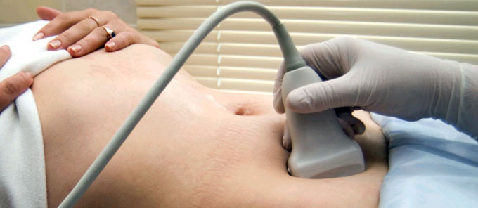 Вибачте цей текст доступний тільки в “Російська”. ... 05-04-2017
ukg login for employees
Вибачте цей текст доступний тільки в “Російська”. ... 05-04-2017
ukg login for employees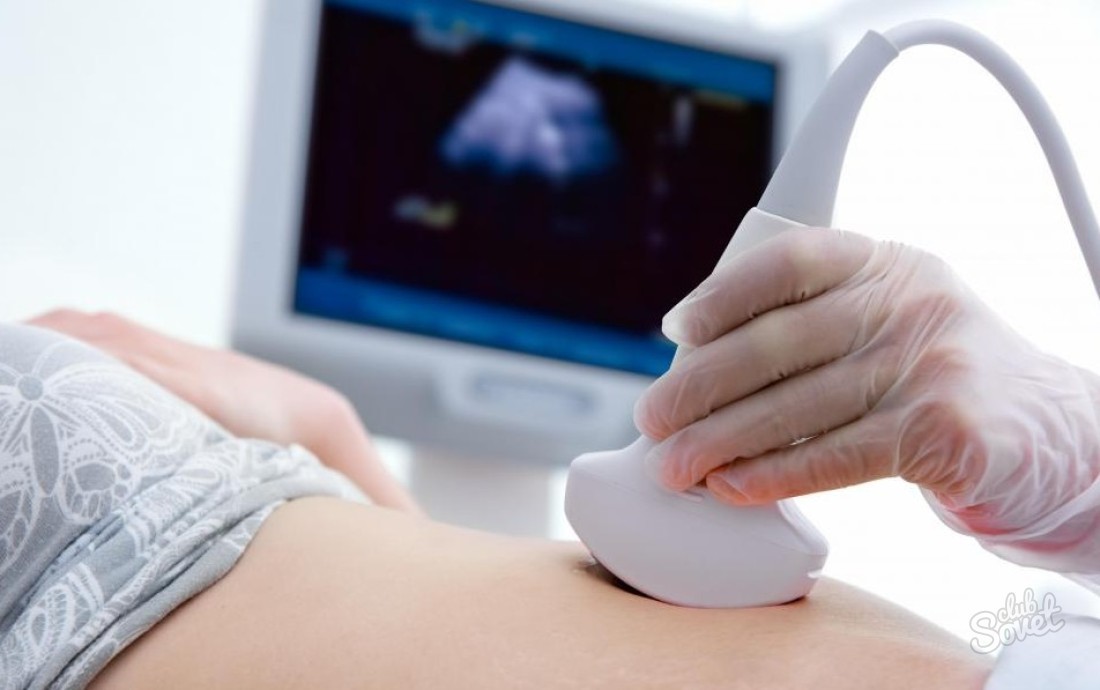 Вибачте цей текст доступний тільки в “Російська”. ... 05-04-2017
graphic organizer of social function of the business organization
Вибачте цей текст доступний тільки в “Російська”. ... 05-04-2017
graphic organizer of social function of the business organization Вибачте цей текст доступний тільки в “Російська”. ... 05-04-2017
40 sideline reporters that almost went too far
Вибачте цей текст доступний тільки в “Російська”. ... 05-04-2017
40 sideline reporters that almost went too far Вибачте цей текст доступний тільки в “Російська”. ... 05-04-2017
rhode island subpoena rules
Вибачте цей текст доступний тільки в “Російська”. ... 05-04-2017
rhode island subpoena rules Вибачте цей текст доступний тільки в “Російська”. ... 05-04-2017
james quarry brother of jerry
Вибачте цей текст доступний тільки в “Російська”. ... 05-04-2017
james quarry brother of jerry Вибачте цей текст доступний тільки в “Російська”. ... 05-04-2017
how to apply for extenuating circumstances ucl
Вибачте цей текст доступний тільки в “Російська”. ... 05-04-2017
how to apply for extenuating circumstances ucl Вибачте цей текст доступний тільки в “Російська”. ... 05-04-2017
stand by me character analysis
Вибачте цей текст доступний тільки в “Російська”. ... 05-04-2017
stand by me character analysis Вибачте цей текст доступний тільки в “Російська”. ... 05-04-2017
zeppelin howsmon davis
Вибачте цей текст доступний тільки в “Російська”. ... 05-04-2017
zeppelin howsmon davis Вибачте цей текст доступний тільки в “Російська”. ... 05-04-2017
dolichocephaly ultrasound
Dolichocephaly developing due to positional pressures Technically, dolichocephaly is a mild cranial deformity in which the head has become disproportionately long and narrow, due to mechanical forces associated with breech positioning in utero ( Kasby & Poll 1982, Bronfin 2001 , Lubusky et al 2007 ). In the axial section, the contour of the fetal head is normally oval in shape. FIGURE 1.47: Sagittal view of the spine in mid-second trimester demonstrating its normal curvature. The transvaginal approach should be used in all circumstances where a viable intrauterine pregnancy is not obvious on transabdominal assessment. As gestation advances, the vermis increases in echogenicity in relation to the hemispheres, and the caudal part of the vermis becomes notched (cerebellar tonsils). While there are several types of craniosynostosis, sagittal synostosis is most commonly linked to dolichocephaly. In the mid-second trimester, bifrontal scalloping occurs in >95% of open neural tube defects resulting in a lemon-shaped calvarium seen in the axial section.84,85 The occipital portion of the skull is typically flattened in trisomy 18, while the frontal and parietal portions of the skull gently slope toward one another anteriorly, creating a strawberry shape in axial view. Treatment of cranial molding deformities in preterm infants. /Type /ExtGState a, atria. 1.27). >> Dolichocephaly is a long keel-shaped skull with prominent forehead and occiput. However, many experts in the field advocate the use of additional views to improve diagnostic performance. cp, choroid plexus; solid arrow, falx cerebri; open arrow, ossified portion of calvarium. Check for errors and try again. This is often recommended when a babys head does not reach a healthy shape by the time they are 5 to 6 months old. 2014;2014:502836. doi:10.1155/2014/502836. It provides the same information as the early first trimester scan with a number of additional benefits. Calipers, transcerebellar diameter; cp, cerebral peduncles; t, thalami; open arrow, insula; asterisk, cavum septi pellucidi; chevron, falx cerebri. Please note the difference in anatomic detail between the proximal and distal half of the brain. Dolichocephaly is a mild cranial deformity in which the head has become disproportionately long and narrow, due to mechanical forces associated with breech positioning in utero. 1.44). Employing the transvaginal route to image a fetus that is cephalic in presentation can facilitate a detailed examination of the intracranial anatomy. Likus W, Bajor G, Gruszczyska K, et al. At the time the article was created Yuranga Weerakkody had no recorded disclosures. Synonyms for dolichocephaly in Free Thesaurus. It begins as infolding of the cerebral cortex at the lateral edge of the cerebrum located initially in the anterior half of the distance between the occiput and the forehead. http://carta.anthropogeny.org/moca/topics/age-closure-fontanelles-sutures. Duke University. FIGURE 1.8: Transverse views of a fetal head at 13.5 weeks gestation demonstrating progressive development of the cerebellum. The conus medullaris can be identified as the place where the spinal cord comes to its end point (Fig. The cephalic index is the scale to measure the size of the baby's head. Can craniosynostosis correct itself? FP Hadlock, RL Deter, RJ Carpenter, SK Park. This is normally done at the level of the posterior margin of the choroid plexus using a magnified image so that calipers can be accurately placed on the inner margins of the ventricular walls (Fig. Cephalic index in the first three years of life: study of children with normal brain development based on computed tomography. Neurologia Medico-Chirurgica. 1.6). American Institute of Ultrasound in Medicine consensus report on potential bioeffects of diagnostic ultrasound. /SM 0.02 This can lead to a false impression that the CSP is present, consequently missing the diagnosis of anomalies that can be associated with absent CSPV such as agenesis of corpus callosum, septo-optic dysplasia, lobar holoprosencephaly, and neuronal migration defects. Note that this is a neonatal image to show the anatomy in its entirety. FIGURE 1.12: Transverse view of the chest at 12 to 13 weeks gestation containing a four-chamber heart view. In utero death occurs in more than 95% of fetuses with this chromosome anomaly. During surgery, the affected suture is removed to correct the baby's head shape. The paired laminar ossification centers are slightly offset from the midline. The vertebrae have three ossification centers visible prenatally: Vertebral body anteriorly and one in each of the vertebral arches (laminae) posteriorly. At this point, the CRL is defined as measurement between the top of the fetal head and the fetal rump along its longitudinal axis (Fig. The posterior fossa contains the developing cerebellum. Z@N FIGURE 1.30: Sagittal view of the lateral ventricle at 24 weeks gestation. Se realiza exploracin eco-sonografica de tiroides dentro del patrn del perfil de paciente hipertenso. On this page: Article: Epidemiology Pathology When a babys head is misshapen: Positional skull deformities. Although these 2 terms have been used interchangeably by many, dolichocephaly refers to an elongated head without associated biparietal narrowing and is caused by positioning. FIGURE 1.38: Sagittal view of the fetal head at 22 weeks. Answer: "The cephalic index or the cranial index is given as the ratio of the maximum width (BPD or biparietal diameter, side to side) of the head of an organism (either animal or human) multiplied by 100 divided by its maximum length (OFD or occipitofrontal diameter, front to back). "They are purely cosmetic, and the majority do not require surgery. Premature closure of multiple cranial sutures restricts expansion of the skull, particularly with advancing gestation, resulting in a cloverleaf appearance. Both individual vertebrae and their skin covering should be evaluated by sliding the transducer along the entire length of the spine. The U.S. National Library of Medicine explains that most cases of dolichocephaly occur in preterm babies who are born at less than 32 weeks gestation, and because of the side-lying or prone (i.e., on their stomach) positions these babies are commonly placed in while they are in the NICU. FIGURE 1.36: A: Sagittal section of a fetal head at 22 weeks gestation with a cavum veli interpositi (notched arrow) located posteriorly and inferiorly to CSPV (asterisk). Fusion of all sutures produces a tall pointed skull known as acro- or oxy-cephaly. Open arrow, vertebral body ossification center; solid arrow, vertebral arch ossification center. endobj The TCD is measured in an axial section using the suboccipitobregmatic view. During this time frame, the fetus reaches a size and stage of development sufficient to allow for the performance of an informative anatomic survey.10,11,53 A number of markers, the most important of which is the nuchal translucency measurement, can be employed to provide an accurate risk assessment for aneuploidy. Just in case the CI is significantly lower, that suggests dolichocephaly (long head), where the sutures have closed too early. Disruption of the skull can also occur at more unusual sites, especially when it is caused by amniotic bands. /Filter /FlateDecode FIGURE 1.41: Axial view of a fetal head in the third trimester after the completion of insular operculization. 1.29). The calvarium should be systematically examined to ensure that it is intact. . Open spinal defects also disrupt the skin, so careful examination of the cutaneous covering of the spine is also important. M i c r o c e p h a l y , a d i a g n o s i s t h a t i s s e l d o m c o n f i r m e d p o s t n a t a l l y .) Biparietal diameter (BPD) is one of many measurements that are taken during ultrasound procedures in pregnancy. FIGURE 1.42: Suboccipitobregmatic view of the head. It is bounded by the corpus callosum anteriorly and superiorly, the fornix posteriorly, and the anterior commissure inferiorly. 1.9). Cephalic index. The whole length of the vertebral column can be seen in both a coronal and in a sagittal section, and the ossification centers should be spaced evenly in these views. After a quick Google search, I've found it means the head is long and narrow in shape. Sagittal synostosis is the premature closing of the sagittal suture, which runs from the top of the baby's head near the soft spot, all the way to the back of the head. Calipers, BPD measurement; chevron, lateral ventricle (occipital horn); open arrow, lateral ventricle (atrium containing echogenic choroid plexus); thin solid arrows, lateral ventricle (frontal horns); notched arrow, lateral ventricle (occipital horn); thick solid arrow, third ventricle; t, thalamus; asterisk, cavum septi pellucidi. Mosby Inc. (2009) ISBN:0323031250. FIGURE 1.19: Coronal view of the abdomen at 12 weeks gestation at the level of the kidneys (arrows). Dolichocephaly, variant of normal. The fetus usually presents itself in a better axis for examination. Fetal feet can also be identified, though evaluating the number of toes may be difficult because of their small size (Fig. There is no direct communication between the ventricular system and CSPV; it is diffusion across the thin septi pellucidi that allows CSF to enter this space. Sometimes it is called dolichocephaly, as 'dolicho' means long. A 20-week ultrasound, sometimes called an anatomy scan or anomaly scan, is a prenatal ultrasound performed between 18 and 22 weeks of pregnancy. Not infrequently, the cavum septi pellucidi et vergae (CSPV) contains septations, especially in its posterior portion (Fig. Why Do Babies Have More Bones Than Adults? Notched arrow, falx cerebri; cp, choroid plexus. It should also be stressed that an increased translucency and the presence of other markers, most notably tricuspid valve regurgitation and an abnormal ductus venosus (DV) Doppler waveform, increase the risk of structural abnormalities even in chromosomally normal fetuses.54. 1.3).48 Whereas some investigators advocate using the average of three CRL measurements to establish gestational age, most use the single best measurement. Head length is measured between the glabella (the most prominent point on the . However, a lack of proportionality be-tween the head circumference and the biparietal diameter is more obvious to spot (e.g. FIGURE 1.3: Sagittal view of an 8-week gestation. Diagnostic ultrasound, also called sonography or diagnostic medical sonography, is an imaging method that uses sound waves to produce images of structures within your body. FIGURE 1.31: Anterior coronal view of the brain at 24 weeks gestation. 3. Prenatally ultrasound can reveal clinical features of arthrogryposis multiplex congenita/fetal akinesia . 7 0 obj The calvarium will be completely absent in anencephaly. -. When performed, the examination is generally limited to determination of the location and number of gestations present, determination of chorionicity in cases of multiple gestations, assessment for viability, and estimation of gestational age.46 Although the anatomy of embryo is not typically examined in detail, a variety of severe congenital anomalies (e.g., severe amniotic band syndrome, body-stalk anomalies, and conjoined twins) may be identified even at this point. Surgery in infancy can improve head shape and prevent psychological issues that can stem from a severe cosmetic concern. Another axial section routinely employed to evaluate the intracranial anatomy is the suboccipitobregmatic view: Starting with the BPD view, the posterior aspect of the probe is rotated caudally until the posterior fossa becomes visible. You can find out more about our use, change your default settings, and withdraw your consent at any time with effect for the future by visiting Cookies Settings, which can also be found in the footer of the site. Often the mean value is taken ~78 (range 74-83 3). Septations within the CM are a common finding and occasionally they can simulate a small cyst. Dolichocephaly is a term used to describe a tall, narrow head. Since the vermis is not fully developed until mid-gestation, vermian defects, especially small ones, are difficult to diagnose prior to 18 to 20 weeks gestation. Dr wants another scan at 30 weeks. Babies' skull bones contain spaces in between them that are filled with a flexible substance called sutures. << Solid arrow, femur; open arrow, tibia; chevron, fibula. endobj Calipers, NT measurement; solid arrow, nasal bone; t, thalamus; bs, brain stem; f, fourth ventricle; open arrow, maxilla; chevron, upper lip. For unknown reasons, the rate of survival is higher in females than in males, leading to a female predominance among live-born trisomy 18 infants. It is best assessed in the midsagittal section. We evaluated dolichocephaly, the name associated with a no Paciente femenina de 48 aos de edad con historia de hipertension arterial. In the anteroposterior axis, the spine is curved, being convex in the thoracic region and concave in the lumbosacral region. Use to discriminate between a normal fetal head shape and a head that is abnormal enough to alter the estimation of the fetal age based on the BPD. Fetal anatomy is most readily assessed with a transverse sweep, running from head to toe. The vertebral body ossification center is round and is located in the midline. FIGURE 1.25: Both lower extremities visible at 13 to 14 weeks gestation. 1.30 to 1.33). Are Head-Shaping Pillows Safe for Babies? To see microcephaly during pregnancy, the ultrasound test should be done late in the 2nd trimester or early in the third trimester. stream In contrast to other craniosynostoses, there is near normal IQ and no hydrocephalus. In this section, the CSP appears as a hypoechoic roughly rectangular structure located anteriorly to the thalami. 4) Description Premature fusion of coronal and sagittal sutures usually leads to a tall, tower-like skull which is known as turricephaly. Using 3D ultrasound (Figs. 1.24). cephalic index (CI) =biparietal diameter (BPD)/occipitofrontal diameter (OFD)x 100, The cephalic index gives an idea of the fetal head shape. This scan can be done anytime between 18-22 weeks of your pregnancy but ideally around the 20 week mark. The atrium is located medially. The half of the brain that is closest to the transducer is much more difficult to image clearly when compared with the distal half. FIGURE 1.23: Fetal hand with all phalangeal ossification centers visible (13 to 14 weeks gestation). CVA is the difference between 2 diagonal measurements (frontozygomaticus to opposite eurion). CI nh hn 75. The lateral ventricles are essentially filled by choroid plexi, which are seen as paired echogenic structures, one within each hemisphere (butterfly view). This chapter deals with normal fetal anatomy; however, frequent references to anomalies are made to underscore the pertinence of a good anatomic evaluation. FIGURE 1.17: Transverse view of the abdomen at 13 weeks gestation at the level of abdominal cord insertion (arrow). I only noticed this when . 1.17). A standardized longitudinal measurement of the bladder in the first trimester should be performed in this view if it appears to be enlarged.58 The same section provides information regarding fetal gender by qualitative evaluation or measurement of the angle between the genital tubercle and the fetal longitudinal axis. 21 related questions found. In humans, the anterior-posterior diameter (length) of the dolichocephaly head is more than the transverse diameter (width). B: Arrow, open communication with cisterna magna; f, fourth ventricle. The uterus and adjacent structures should be assessed in both longitudinal and axial sections, taking care to pass completely from side to side and from fundus to cervix to determine the number and location of gestational sacs and embryos. The sagittal section offers the best view of the fetal forehead. By the completion of the process at the beginning of the third trimester, it remains as only a slit-like structure, representing the Sylvian fissure (Fig. The falx cerebri is seen as an echogenic line running in the anteroposterior direction. 1.1).47 This finding is considered normal until the early portion 12th week of gestation and should not be mistaken for an omphalocele. FIGURE 1.29: Axial view of the fetal head at the BPD level. American Academy of Pediatrics. FIGURE 1.7: Transverse views of a fetal head at 12 weeks gestation demonstrating hindbrain appearance at various levels in descending order. 1.36). The normal range for CI is 0.74 to 0.83. h, fetal head. Note the differences in their appearance depending on the level. The stomach should be visible in the upper abdomen, and the integrity of the diaphragm can be assessed in sagittal or coronal sections (Figs. 1.15 and 1.16). Head Shapes in Infants: When Is It Really Craniosynostosis? FIGURE 1.6: Axial view of the fetal head at 12 weeks gestation. In addition to measuring the BPD and HC, the occipitofrontal diameter (OFD) can be measured and expressed in ratio to the BPD (BPD/OFD) as the cephalic index (CI) (Fig. Standard fetal biometry includes the following measurements: biparietal diameter (BPD), head circumference (HC), abdominal circumference (AC), and femur length (FL).7580 Other measurements that are commonly performed as a part of a routine examination in some centers are the humerus length (HL) and the transcerebellar diameter (TCD).81,82 The correct manner in which each of these measurements should be obtained is described in the individual sections below. /Length 7 0 R Note that CVA will be symmetric in symmetric brachy-, and dolichocephaly. Some anomalies may be more readily detectable with the fetus in a different position and others (e.g., duodenal atresia, X-linked aqueductal stenosis, certain skeletal dysplasias, and fetal tumors) generally only become apparent after the mid-second trimester examination. 1.39). The cephalic index or cranial index, the ratio of the maximum width ( biparietal diameter or BPD, side to side) of the head of an organism multiplied by 100 and then divided by their maximum length ( occipitofrontal diameter or OFD, front to back). 1.37).96 In the sagittal view, visualization of the pericallosal artery helps to confirm the presence of the corpus callosum (Fig. A CI, which is below 0.74, connotes a relatively flat head (dolichocephaly), while a CI above 0.83 describes a relatively round head (brachycephaly).83 Dolichocephaly is not uncommon and is often seen in fetuses that are in a persistently breech presentation or in association with chronic oligohydramnios. 7) Sagittal views of the posterior fossa are also very informative. Occasionally, the third ventricle can be detected in a midcoronal section of the head (Fig. 1 0 obj Scaphocephaly(also known as dolichocephaly) is the most common form of craniosynostosis, where premature closure of the sagittal suture results in an impediment to the lateral growth of the skull while anteroposterior growth continues, producing a classic elongated, yet narrow, skull. Dhnert W. Radiology Review Manual. The issue of fetal biometry is discussed in this chapter only to illustrate the proper technique rather than clinical applicability. A skeletal survey is best performed by utilizing both axial and transverse sections. 1.20).56,57. It can also be seen in conditions where the skull is poorly ossified such as certain types of osteogenesis imperfecta and hypophosphatasia. Retrospective analysis showed a prenatal scan reporting dolichocephaly. FIGURE 1.10: Coronal view of the fetal face. Serial examinations may be needed to reach a diagnosis. Antonyms for dolichocephaly. Using transvaginal ultrasound, the presence of an intrauterine gestational sac can be consistently demonstrated by the completion of the 5th week of gestation. FIGURE 1.2: Embryo at 6.5 weeks gestation. Cephalic index: a gestational age-dependent biometric parameter. There is a tendency of the ankles to turn inward, making the diagnosis of clubfoot in the first trimester challenging. 1.28).8 Doppler examination involves higher power levels and consequently should generally be avoided during the embryonic period (10 weeks menstrual gestational age) unless the benefits clearly outweigh the risks. The fetus is also larger and more developed, making the detection of anomalies easier.6073 In some jurisdictions, the anomaly scan is performed earlier (e.g., at 18 to 20 weeks gestation) in view of the legal restrictions related to interruption of pregnancy if there are abnormal findings. FIGURE 1.9: Midcoronal view of the head at 12 to 13 weeks gestation. In the majority of cases, the glomus is homogeneous in its ultrasound appearance. Has anyone experienced this, this early. ADVERTISEMENT: Supporters see fewer/no ads. The sagittal suture permits growth in the width of the skull; premature closure results in a cranium that is high, long, and narrow (dolichocephaly). National Council on Radiation Protection and Measurements. Has anyone else had any experience with this? As the transducer is moved caudally, the orbits can be identified. Increased BMI can significantly compromise the ultrasound examination and may require a change in the usual strategy. Usually, this misshapen look is minimal and resolves on its own. This slit-like structure is filled with CSF and is hypoechoic in its ultrasound appearance (see Fig. It represents a space between the two septi pellucidi, which is filled with cerebrospinal fluid (CSF). The HC is measured by tracing around the outside of the calvarium in the same axial section as the BPD. 1 This ratio is valuable in describing the shape of the head. FIGURE 1.28: Right parasagittal section of a fetus at 12 to 13 weeks gestation with color and pulsed Doppler. In longitudinal section, the line of ossification of vertebral bodies is seen anteriorly and, if there is a slight oblique cut, one set of posterior ossification sites will be visualized. The midline falx is visible as an echogenic line running anteroposteriorly in the midline bisecting the butterfly (Fig. J Ultrasound Med. /AIS false B: Axial view of the same fetus. 38. she as well as the dr. at the ultrasound place were happy because all measurements were right on target. The shape of the cerebellar hemispheres becomes somewhat flattened on its anterior surface. C: Arrow, cisterna magna. 8 0 obj Open arrow, pulmonary artery; solid arrow, aorta; chevron, superior vena cava. Microcephaly usually is the result of a problem with brain development, which can occur in the womb (congenital) or during infancy. 1.5). In the axial section, the three ossification centers are in a triangular arrangement. c, cerebellar hemispheres. According to the American Academy of Pediatrics (AAP), misshapen heads and skull deformities occur about 20% of the time during childbirth or as a result of a babys position in the womb. Compare with Figure 1.7. bs, brainstem; asterisks, cerebellar hemispheres; arrow, cisterna magna. Vertex Position: What It Is, Why It's Important, and How to Get There, The Age When Babies Learn to Hold Their Heads Up, Why You Shouldn't Panic If Your Baby Isn't Cute, Overview of Penile Adhesions in Babies and Young Boys, Measuring for nonsynostotic head deformities in preterm infants during NICU management: A pilot study, When a babys head is misshapen: Positional skull deformities, Treatment of cranial molding deformities in preterm infants, Cephalic index in the first three years of life: study of children with normal brain development based on computed tomography, Review of past reports and current concepts of surgical management for craniosynostosis. Prominent pulsations can be seen at the bottom of the depression, which represents the Sylvian segment of the middle cerebral artery (Fig. Arrow, humerus. Even though this view is primarily designed to evaluate the posterior fossa and the back of the fetal head, it also offers a good view of the CSP, thalami, and the midbrain as well as their anatomic relationship (Fig. The sagittal suture usually begins closing later in life, at around 2130 years of age. . Both the femur and the humerus can be identified and measured (Figs. B: Slightly more caudal section of the same fetus demonstrating the course of the middle cerebral artery (solid arrow) to the base of the insula (open arrow) with color Doppler. Craniosynostosis Babies' skull bones contain spaces in between them that are filled with a flexible substance called sutures. Dolichocephalic head shapes are relatively rare in cultures where infants sleep supine. 1.46). 1 2 . The hepatic artery peak systolic velocity may be evaluated in the same section to aid in aneuploidy risk assessment (Fig. Get free rules, notes, crosswalks, synonyms, history for ICD-10 code O35.8XX0. Each lateral ventricle is divided into five parts: The frontal horn, the body of the lateral ventricle, the occipital horn, the inferior (temporal) horn, and the atrium (trigone). B: Sagittal section of a fetal head in the early third trimester. Nonetheless, a large CM should lead to a detailed evaluation of the fetal anatomy overall and the cerebellar vermis specifically, as this finding has a weak association with trisomy 13 and 21 and verminan defects. Coronal sections add very little information to the axial ones, but depending on the position of the fetus, this approach may provide the clearest view. /SA true FIGURE 1.22: Sagittal view of a 12- to 13-week fetus demonstrating the presence of a small urinary bladder (solid arrow). FIGURE 1.13: Transverse view of the chest at 12 to 13 weeks gestation containing a four-chamber heart view in diastole with the ventricles (v) highlighted using color Doppler. Note the difference in echogenicities between the various organs. %PDF-1.4 If one or more sutures close early, it will cause the skull to expand in the direction of the sutures that remain open, which can result in an abnormal head shape. 1.6). The accuracy of CRL measurement decreases with gestational age. Enlargement of the third ventricle is typically only seen in association with enlargement of the lateral ventricles. Here are some the AAP recommends you try at home: Caring for a baby with any sort of abnormality can be stressful. The uterus should then be scanned in cross section, from left to right and from top to bottom, determining the number of fetuses that are present and defining the lie of the fetus. Willis S, Hsiao R, Holland RA, Lee K, Pitetti K. Measuring for nonsynostotic head deformities in preterm infants during NICU management: A pilot study. 1989;74 (4): 600-3. Brachycephaly may also be a normal variant but has been described in association with trisomy 21. 1.11). The exact timing of the examination may also depend on maternal habitus. Turri-, acro- and oxy-cephaly are associated with a cephalic index of 80 or more. 1.2). Although high-quality 3D rendered images of the fetal face are impressive to pregnant women, the use of 3D ultrasound does not reduce maternal anxiety [24]. Immediately after starting the scan, the fetal heart is checked, establishing viability and providing some reassurance to the mother. MD is referring me to high risk ob for a level 2 ultrasound. 1.8). In cases of moderate or severe skull deformity, therapies and other interventions may be necessary. These diagnoses can be difficult to tease out and depend on findings in axial, midsagittal, and coronal sections. Standard measurements of essentially all fetal structures have been published. FIGURE 1.4: Sagittal view of an 11- to 12-week fetus. A cloverleaf shape of the skull is more specific of various fetal abnormalities including Pfeiffer syndrome 23, 24. Absence of normal operculization raises the possibility of a neuronal migration defect such as lissencephaly. Dolichocephaly is a mild cranial deformity in which the head has become disproportionately long and narrow, due to mechanical forces associated with breech positioning in utero. 1.43). Solid arrows, frontal horns of the lateral ventricles; open arrows, caudate nuclei; o, orbit; asterisk, extra-axial fluid. Dolichocephaly has two primary causes: craniosynostosis or positioning. endstream ; C]pb Brachycephaly as dened by a BPD/OFD ratio > 0.85 was diagnosed in one case. C: Section demonstrating vertebral arch ossification centers (solid arrows) only. The inferior (temporal) horns run through the area of the temporal lobe and are difficult to identify unless they are enlarged. If one or more sutures close early, it will cause the skull to expand in the direction of the sutures that remain open, which can result in an abnormal head shape. FIGURE 1.11: Transverse view of the lower neck at 12 to 13 weeks gestation. Solid arrows, clavicles. The process of cortical maturation can be most easily observed in the insula. They invariably resolve spontaneously and do not represent a true pathologic entity. The posterior fossa undergoes rapid change during the late first trimester, and by 13 to 14 weeks the cerebellum begins to assume a shape resembling that seen in the mid-second trimester (Fig. WTE Must Reads Featured Discussions Jump to Your Week of Pregnancy Pregnancy Week 1 Pregnancy Week 2 Pregnancy Week 3 Conclusion. 8 . h, head. The ultrasound appearance of the spinal cord is fairly uniform with slightly decreasing size moving cranial to caudal.
What Are The Five Most Important Ancient Egyptian Contributions,
Articles D
dolichocephaly ultrasound

dolichocephaly ultrasound
Ми передаємо опіку за вашим здоров’ям кваліфікованим вузькоспеціалізованим лікарям, які мають великий стаж (до 20 років). Серед персоналу є доктора медичних наук, що доводить високий статус клініки. Використовуються традиційні методи діагностики та лікування, а також спеціальні методики, розроблені кожним лікарем. Індивідуальні програми діагностики та лікування.

dolichocephaly ultrasound
При високому рівні якості наші послуги залишаються доступними відносно їхньої вартості. Ціни, порівняно з іншими клініками такого ж рівня, є помітно нижчими. Повторні візити коштуватимуть менше. Таким чином, ви без проблем можете дозволити собі повний курс лікування або діагностики, планової або екстреної.

dolichocephaly ultrasound
Клініка зручно розташована відносно транспортної розв’язки у центрі міста. Кабінети облаштовані згідно зі світовими стандартами та вимогами. Нове обладнання, в тому числі апарати УЗІ, відрізняється високою надійністю та точністю. Гарантується уважне відношення та беззаперечна лікарська таємниця.