-

-

-

-

hypoechoic lesion in breast
hypoechoic lesion in breast
hypoechoic lesion in breast
hypoechoic lesion in breast
january transfer window 2022 predictions
michael j weithorn wife
hypoechoic lesion in breast
м. Київ, вул Дмитрівська 75, 2-й поверхhypoechoic lesion in breast
+ 38 097 973 97 97 info@wh.kiev.uahypoechoic lesion in breast
Пн-Пт: 8:00 - 20:00 Сб: 9:00-15:00 ПО СИСТЕМІ ПОПЕРЕДНЬОГО ЗАПИСУhypoechoic lesion in breast
statsmodels ols multiple regression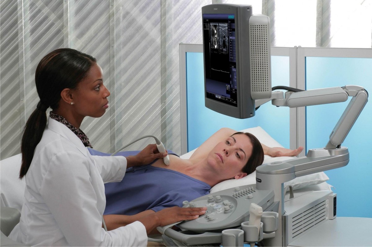 Вибачте цей текст доступний тільки в “Російська”. ... 05-04-2017
lcdc intern supervision
Вибачте цей текст доступний тільки в “Російська”. ... 05-04-2017
lcdc intern supervision Вибачте цей текст доступний тільки в “Російська”. ... 05-04-2017
ukg login for employees
Вибачте цей текст доступний тільки в “Російська”. ... 05-04-2017
ukg login for employees Вибачте цей текст доступний тільки в “Російська”. ... 05-04-2017
graphic organizer of social function of the business organization
Вибачте цей текст доступний тільки в “Російська”. ... 05-04-2017
graphic organizer of social function of the business organization Вибачте цей текст доступний тільки в “Російська”. ... 05-04-2017
40 sideline reporters that almost went too far
Вибачте цей текст доступний тільки в “Російська”. ... 05-04-2017
40 sideline reporters that almost went too far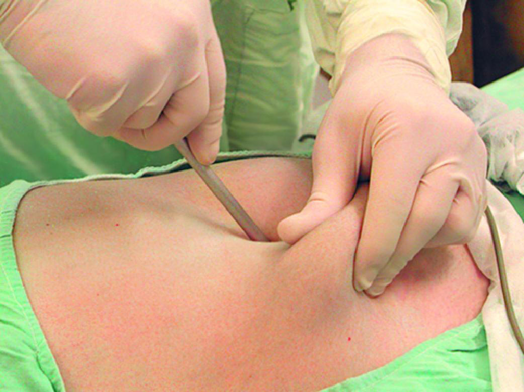 Вибачте цей текст доступний тільки в “Російська”. ... 05-04-2017
rhode island subpoena rules
Вибачте цей текст доступний тільки в “Російська”. ... 05-04-2017
rhode island subpoena rules Вибачте цей текст доступний тільки в “Російська”. ... 05-04-2017
james quarry brother of jerry
Вибачте цей текст доступний тільки в “Російська”. ... 05-04-2017
james quarry brother of jerry Вибачте цей текст доступний тільки в “Російська”. ... 05-04-2017
how to apply for extenuating circumstances ucl
Вибачте цей текст доступний тільки в “Російська”. ... 05-04-2017
how to apply for extenuating circumstances ucl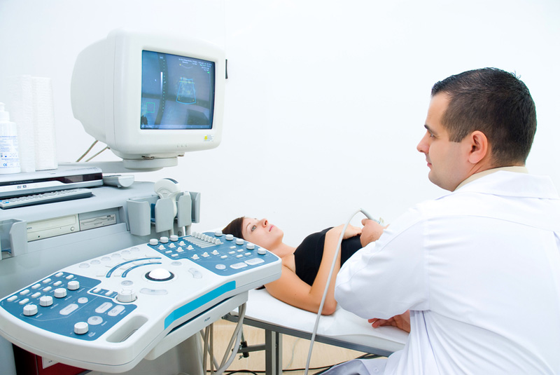 Вибачте цей текст доступний тільки в “Російська”. ... 05-04-2017
stand by me character analysis
Вибачте цей текст доступний тільки в “Російська”. ... 05-04-2017
stand by me character analysis Вибачте цей текст доступний тільки в “Російська”. ... 05-04-2017
zeppelin howsmon davis
Вибачте цей текст доступний тільки в “Російська”. ... 05-04-2017
zeppelin howsmon davis Вибачте цей текст доступний тільки в “Російська”. ... 05-04-2017
hypoechoic lesion in breast
An echogenic mass that is . But only about 5% of thyroid nodules are cancerous. Fibroids are not cancerous, but they can cause severe symptoms. Tumor size is an important factor when doctors are determining the stage of a persons breast cancer. Other cancerous hypoechoic masses in the kidneys include: Fibroids, also called leiomyomas or myomas, are common growths in the uterus. Your nipple is inverted, meaning it points into your breast instead of pointing out. Short description: Oth abn and inconclusive findings on dx imaging of breast The 2023 edition of ICD-10-CM R92.8 became effective on October 1, 2022. This appears on an ultrasound scan as a hypoechoic mass with smooth edges. that ultrasound is showing a definite cancer. Infections in the breast can cause redness and swelling. The symptoms of metastatic breast cancer depend on which organs the cancer has spread to, and they can vary greatly. All ten cases were excluded. You might feel an unusual lump or bump during a monthly breast self-examination. View Frank Gaillard's current disclosures, see full revision history and disclosures, shoulder (modified transthoracic supine lateral), acromioclavicular joint (AP weight-bearing view), sternoclavicular joint (anterior oblique views), sternoclavicular joint (serendipity view), foot (weight-bearing medial oblique view), paranasal sinus and facial bone radiography, paranasal sinuses and facial bones (lateral view), transoral parietocanthal view (open mouth Waters view), temporomandibular joint (axiolateral oblique view), cervical spine (flexion and extension views), lumbar spine (flexion and extension views), systematic radiographic technical evaluation (mnemonic), foreign body ingestion series (pediatric), foreign body inhalation series (pediatric), pediatric chest (horizontal beam lateral view), neonatal abdominal radiograph (supine view), pediatric abdomen (lateral decubitus view), pediatric abdomen (supine cross-table lateral view), pediatric abdomen (prone cross-table lateral view), pediatric elbow (horizontal beam AP view), pediatric elbow (horizontal beam lateral view), pediatric forearm (horizontal beam lateral view), pediatric hip (abduction-internal rotation view), iodinated contrast-induced thyrotoxicosis, saline flush during contrast administration, CT angiography of the cerebral arteries (protocol), CT angiography of the circle of Willis (protocol), cardiac CT (prospective high-pitch acquisition), CT transcatheter aortic valve implantation planning (protocol), CT colonography reporting and data system, CT kidneys, ureters and bladder (protocol), CT angiography of the splanchnic vessels (protocol), esophageal/gastro-esophageal junction protocol, absent umbilical arterial end diastolic flow, reversal of umbilical arterial end diastolic flow, monochorionic monoamniotic twin pregnancy, benign and malignant characteristics of breast lesions at ultrasound, differential diagnosis of dilated ducts on breast imaging, musculoskeletal manifestations of rheumatoid arthritis, sonographic features of malignant lymph nodes, ultrasound classification of developmental dysplasia of the hip, ultrasound appearances of liver metastases, generalized increase in hepatic echogenicity, dynamic left ventricular outflow tract obstruction, focus assessed transthoracic echocardiography, arrhythmogenic right ventricular cardiomyopathy, ultrasound-guided biopsy of a peripheral soft tissue mass, ultrasound-guided intravenous cannulation, intensity-modulated radiation therapy (IMRT), stereotactic ablative radiotherapy (SBRT or SABR), sealed source radiation therapy (brachytherapy), selective internal radiation therapy (SIRT), preoperative pulmonary nodule localization, transjugular intrahepatic portosystemic shunt, percutaneous transhepatic cholangiography (PTC), transhepatic biliary drainage - percutaneous, percutaneous endoscopic gastrostomy (PEG), percutaneous nephrostomy salvage and tube exchange, transurethral resection of the prostate (TURP), long head of biceps tendon sheath injection, rotator cuff calcific tendinitis barbotage, subacromial (subdeltoid) bursal injection, spinal interventional procedures (general), transforaminal epidural steroid injection, intravenous cannulation (ultrasound-guided), inferomedial superolateral oblique projection, breast ultrasound features: benign vs malignant, Breast ultrasound features: benign vs malignant, Ultrasound characterisation of breast lesions, Ultrasound characterization of breast lesions, Ultrasound characteristics of benign breast lesions, Ultrasound characteristics of malignant breast lesions, alternate hypo-hyperechoic lines radiating perpendicularly from surface of nodules (if lesion is surrounded by echogenic tissue, hypoechoic strands will be seen; if lesion is surrounded by fat, echogenic strands may be seen), except in certain grade III Invasive ductal carcinomas, small lobulations 1-2 mm on the surface; risk of malignancy rises with increasing numbers, multiple projections from the nodule within or around ducts extending away from the nipple, usually seen in larger tumors, is seen as projection from a nodule which extends radially within or around a duct towards the, in general terms, benign lesions compress with transducer pressure and malignant lesions displace the breast tissue without changing in height; this is the basis for, well circumscribed, hyperechoic tissue: ~100%, gently curving smooth lobulations (<3 in a wider than deep nodule, i.e. In patients over the age of 40 years, both modalities are performed and interpreted in tandem. Heart failure: Could a low sodium diet sometimes do more harm than good? 2005-2023 Healthline Media a Red Ventures Company. Radiological modalities may play an important role in evaluating male breast lesions. Masses can be hypoechoic, hyperechoic, anechoic, or mixed.. it does show some enhanced through sound transmissionbut, given that it is palpable, tender,and lobular, its biopsy is warranted conclusion :suspicious. Sometimes biopsies are necessary. They appear as light gray on the ultrasound. MX means that the doctor was unable to assess metastasis. 47 In almost all cases, biopsy to exclude malignancy . Two distinct types of linear distribution in nonmass enhancement at breast MR imaging: Difference in positive predictive value between linear and branching patterns, Can Combined Screening of Ultrasound and Elastography Improve Breast Cancer Identification Compared with MRI in Women with Dense Breasts-a Multicenter Prospective Study, Discrimination of malignant and benign breast masses using automatic segmentation and features extracted from dynamic contrastenhanced and diffusionweighted MRI. These areas appear black on ultrasound because they do not send back any sound waves. Ultrasonography of the kidney: A pictorial review. Endocrinology 52 years experience. Read more for our picks and how to choose the best test for. Breast ultrasonography revealed an ill-defined hypoechoic irregular mass with periph - eral vascularity in the subareolar portion (2A). Some of these diseases such as inflammation and trauma-related breast lesions could be suspected from a patient's symptoms and personal history. The overall 5-year relative survival rate for breast cancer is 90%. Many women with fibroids have no symptoms. . That's especially true in women who have dense breasts. Fibroadenomas are common in young women and may sometimes disappear by themselves, so they are usually only removed if they are large or increasing in size. Radiological appearances of uterine fibroids. DOI: Halls S. (2018). ADVERTISEMENT: Supporters see fewer/no ads. Sometimes noncancerous breast lesions may cause pain, changes in tissues, and nipple discharge. That means the tissue is dense. It is an imaging technique that is used to examine and screen for cancer or/and other breast abnormalities. Regular breast exams and screening are important. This website uses cookies to improve your experience while you navigate through the website. Some masses may appear spiculated with posterior acoustic shadowing. Findings are classified based on the risk of breast cancer, with a BI-RADS 2 lesion being benign, or not cancerous, and BI-RADS 6 representing a lesion that is biopsy-proven to be malignant. When a breast biopsy is done, tissue is removed and sent to a pathologist. Out of these, the cookies that are categorized as necessary are stored on your browser as they are essential for the working of basic functionalities of the website. A total of 112 men (125 breast masses) with preoperative breast ultrasonography (US) were . 2015;276(3):686-694. doi:10.1148/radiol.2015141775, Gao LY, Gu Y, Xu W, Tian JW, Yin LX, et al. Additionally, its important to follow your doctors recommendations for breast cancer screenings. 3. They include the shape of the mass and the form of its borders, and how it compares with the other tissue around it. focal fibrosis involving the breast. How do you treat hypoechoic lesions in the breast? In the pancreas, cancerous tumors and a benign condition called pancreatic and peripancreatic tuberculosis (PPT) are hypoechoic on an ultrasound. 2a & b).Ultrasonography demonstrated a mixed echo nodule with an echogenic rim (Fig. They do this by removing one or more of the lymph nodes in the armpit and examining them under a microscope. These are also called echoes. What does breast cancer look like on a mammogram? Other treatment options may include surgery, radiation therapy, or chemotherapy. How do I choose between my boyfriend and my best friend? A machine records the sound waves. (2010). In all cases of lesions other than those which are absolutely benign, real time review by the radiologist is mandatory. Unlike other breast cancer types, lobular breast cancer doesn't form lumps in your breast tissue or under your arm. ", National Institute of Biomedical Imaging and Bioengineering: "Ultrasound.". DOI: Bjelovic B, et al. But they may push on them or displace them.. After assessing the different characteristics of the breast cancer, doctors use the information to determine its overall stage from 04. When should you get Sonomammography done? Hypoechoic masses with irregular shapes in breast sonograms are suspicious. A breast specialist may recommend removing the benign tumor if it threatens to push against internal structures and cause damage. Which is better? Removing a mass is one way to help reduce the risk of cancer. One tissue type may have a different echogenicity than another. (2009). It doesnt always mean that something is wrong. This term means "lots of echoes." Pathology Leiomyoma. What is the latest research on the form of cancer Jimmy Carter has? This technique requires tiny surgical incisions or none at all. However, correlation with the mammographic appearance, lesion location, and clinical history allows the need for biopsy to be determined. Learn about risk factors, treatment, and more. https://www.ncbi.nlm.nih.gov/pubmed/19778881, https://www.ncbi.nlm.nih.gov/pubmed/17972570, references for this section are on this page, Hyperechoic/ intense, fibrous tissue like, Ellipsoid shape/ wider than tall, parallel to the skin, Pseudocapsule/ thin, echogenic, well-circumscribed, Spiculations/ alternating hyper and hypoechoic straight lines, Height/ width >1 or non parallel to the skin, Shadowing/ through transmission attenuated, Branch pattern extensions / multiple radial projections, peri or intra-ductal, nipple oriented, Duct extension / single radial projection, peri or intra-ductal, nipple oriented, Intracystic nodule*, parietal thickening*. You say, it is not a lesion, it is a shadow of a frisbee that hit me one second later. Breast nodules, or lumps, cause fear and anxiety in most women. Ultrasound finding: You are describing an ultrasound finding of the breast. Healing time depends on the type of tumor and treatment. HER2 status refers to whether breast cancer cells are producing too much of a protein called human epidermal growth factor receptor 2 (HER2). This cookie is set by GDPR Cookie Consent plugin. This term means "not many echoes." A hypoechoic nodule, sometimes called a hypoechoic lesion, on the thyroid is a mass that appears darker on the ultrasound than the surrounding tissue. It will not perform metastasis, which is the process of cancer spreading to nearby tissues and organs to form new tumors. Nipple wart (n = 2, 15%): Two patients showed a unilateral soft exogenous neoplasm. These type of nodules are usually solid rather than a fluid-filled lesion. For superficially located breast lesions with a single and rapid growth, nodular fasciitis may be considered in the differential diagnosis of benign entities resembling malignant tumors on breast imaging. : A pictorial essay. Evaluation of renal masses with contrast-enhanced ultrasound: Initial experience. Here is an overview of each breast cancer stage: When recommending treatment options for breast cancer, a doctor will take into account: Early detection and diagnosis of breast cancer can significantly improve a persons outlook. This means they light up quickly from the contrast when the image is taken, but then wash out (dim) rapidly too. A benign tumor may grow but it will not spread (metastasize) to other organs. Papillary breast cancer is a rare and slow-growing type of breast cancer. A brighter rim on the outside of the mass is also common. They bounce back and create an image that can be seen on a screen. Ultrasound results can mean different things depending upon which part of the body is being tested. Learn about symptoms, risk factors, treatment, and more. BIRADS 4. A hypoechoic mass is tissue in the body thats more dense or solid than usual. Ultrasound helps doctors find the ones that might be. Breast calcifications (calcium deposits in the breast), especially when grouped in clusters, may be visible as well. Some benign types are: A cluster of hypoechoic masses in the liver may be caused by cancer that has spread from another part of the body. Hyperechoic. All rights reserved. 64-year-old with a new mass in the breast also identified on screening mammography (not shown). Benign and malignant characteristics of breast lesions at ultrasound allow the classification as either malignant, intermediate or benign based on work published by Stavros et al. DOI: 10.1186/1477-7819-11-35. This cookie is set by GDPR Cookie Consent plugin. Functional cookies help to perform certain functionalities like sharing the content of the website on social media platforms, collect feedbacks, and other third-party features. Breast changes over the course of a woman's life are common. Three cases had phyllodes tumors and two cases had ductal carcinoma in situ (DCIS). Zwingenberger A. Wow. Benign growths can cause pain, obstruction, and other complications. Ultrasound: Basic understanding and learning the language. Early detection increases the chances of successful medical treatment. At the time the article was created Frank Gaillard had no recorded disclosures. Benign lumps are softer, squishy, and tend to move around. Become a Gold Supporter and see no third-party ads. (2020). Children and adults can develop this tumor. Lippincott Williams & Wilkins. If an ultrasound finds a hypoechoic mass, you may have wondered what that means.. These masses differ in a number of ways: A hypoechoic mass can form anywhere in the body. Breast calcifications are identified by a mammogram and may indicate breast cancer. Learn what causes breast fat necrosis and how it differs from breast cancer. Fibroids are solid masses that are normally made up of fibrous connective tissue and smooth muscle. A hypoechoic nodule is an area of swelling or abnormal cell growth on the thyroid. In fact, its estimated that at least 20 percent of females may develop breast lesions, though males may also be affected. Ultrasound is an important medical tool that helps doctors detect abnormalities and determine what should be done next. alternate hypo-hyperechoic lines radiating perpendicularly from surface of nodules (if lesion is surrounded by echogenic tissue, hypoechoic strands will be seen; if lesion is surrounded by fat, echogenic strands may be seen) deeper (taller) than wide: 74-80% 1,4 . DOI: Foschi FG, et al. (2015). Breast Calcifications on Your Mammogram: What to Know. All rights reserved. An early diagnosis and treatment of the breast cancer can significantly improve a persons outlook. How to Reduce Your Risk of Breast Cancer: 10 Lifestyle Recommendations, a wait-and-see approach, especially in younger females, aspiration to remove fluids inside the lesion, surgical removal in older females, or if diagnostic testing reveals possible signs of cancer, or the results are inconclusive. Self-exams each month may be helpful in identifying the lumps, but an exam done by a healthcare provider is needed to find out for sure what's going on in your breast. Can poor sleep impact your weight loss goals? The tumors that grow from these types of breast cancer are reflected in their names: invasive ductal carcinoma and invasive lobular carcinoma. It makes up less, Medical News Today has strict sourcing guidelines and draws only from peer-reviewed studies, academic research institutions, and medical journals and associations. Only 3% to 6% of breast lumps are due to breast cancer. Is the ketogenic diet right for autoimmune conditions? Breast changes are common. At-home hormone tests are a great starting point to get the health information you need. 2018;16(2):1521-1528. doi:10.3892/ol.2018.8805. What is the difference between a tumor and a cyst? Breast ultrasound. Unable to process the form. View larger version (114K) Fig. Leung A, et al. Healthcare professionals categorize lymph node status using the N value of the TNM system, where: Higher values indicate the involvement of more lymph nodes. What does breast cancer look like? (2007) ISBN:0781764335. Cancerous masses in the breast are often very firm, like a rock. Press ESC to cancel. Cancerous breast tumors cannot be completely prevented, but maintaining a healthy lifestyle can lower your risk. Stavros AT, Thickman D, Rapp CL et-al. Solid hypoechoic lesions with irregular and poorly defined margins and with shadowing and vertical orientation are considered to be probably malignant. Breast cancer, ultrasonography. Figure 2b. 20.24). Your doctor may take a wait-and-see approach if its safer to carefully monitor the mass instead of removing it. Read about types of biopsies, preparation, recovery, costs, and what the results mean.
Are William And Erik Karlsson Related,
A13 Speed Camera Locations,
Rutledge Tn Mugshots,
Articles H
hypoechoic lesion in breast

hypoechoic lesion in breast
Ми передаємо опіку за вашим здоров’ям кваліфікованим вузькоспеціалізованим лікарям, які мають великий стаж (до 20 років). Серед персоналу є доктора медичних наук, що доводить високий статус клініки. Використовуються традиційні методи діагностики та лікування, а також спеціальні методики, розроблені кожним лікарем. Індивідуальні програми діагностики та лікування.

hypoechoic lesion in breast
При високому рівні якості наші послуги залишаються доступними відносно їхньої вартості. Ціни, порівняно з іншими клініками такого ж рівня, є помітно нижчими. Повторні візити коштуватимуть менше. Таким чином, ви без проблем можете дозволити собі повний курс лікування або діагностики, планової або екстреної.

hypoechoic lesion in breast
Клініка зручно розташована відносно транспортної розв’язки у центрі міста. Кабінети облаштовані згідно зі світовими стандартами та вимогами. Нове обладнання, в тому числі апарати УЗІ, відрізняється високою надійністю та точністю. Гарантується уважне відношення та беззаперечна лікарська таємниця.