-

bright white spots on mri lumbar spine
bright white spots on mri lumbar spine
why does noel gugliemi always plays hector -

-

-

bright white spots on mri lumbar spine
bright white spots on mri lumbar spine
bright white spots on mri lumbar spine
bright white spots on mri lumbar spine
does takiya like kobayashi
fenwick house ballina
bright white spots on mri lumbar spine
м. Київ, вул Дмитрівська 75, 2-й поверхbright white spots on mri lumbar spine
+ 38 097 973 97 97 info@wh.kiev.uabright white spots on mri lumbar spine
Пн-Пт: 8:00 - 20:00 Сб: 9:00-15:00 ПО СИСТЕМІ ПОПЕРЕДНЬОГО ЗАПИСУbright white spots on mri lumbar spine
txt comeback countdown live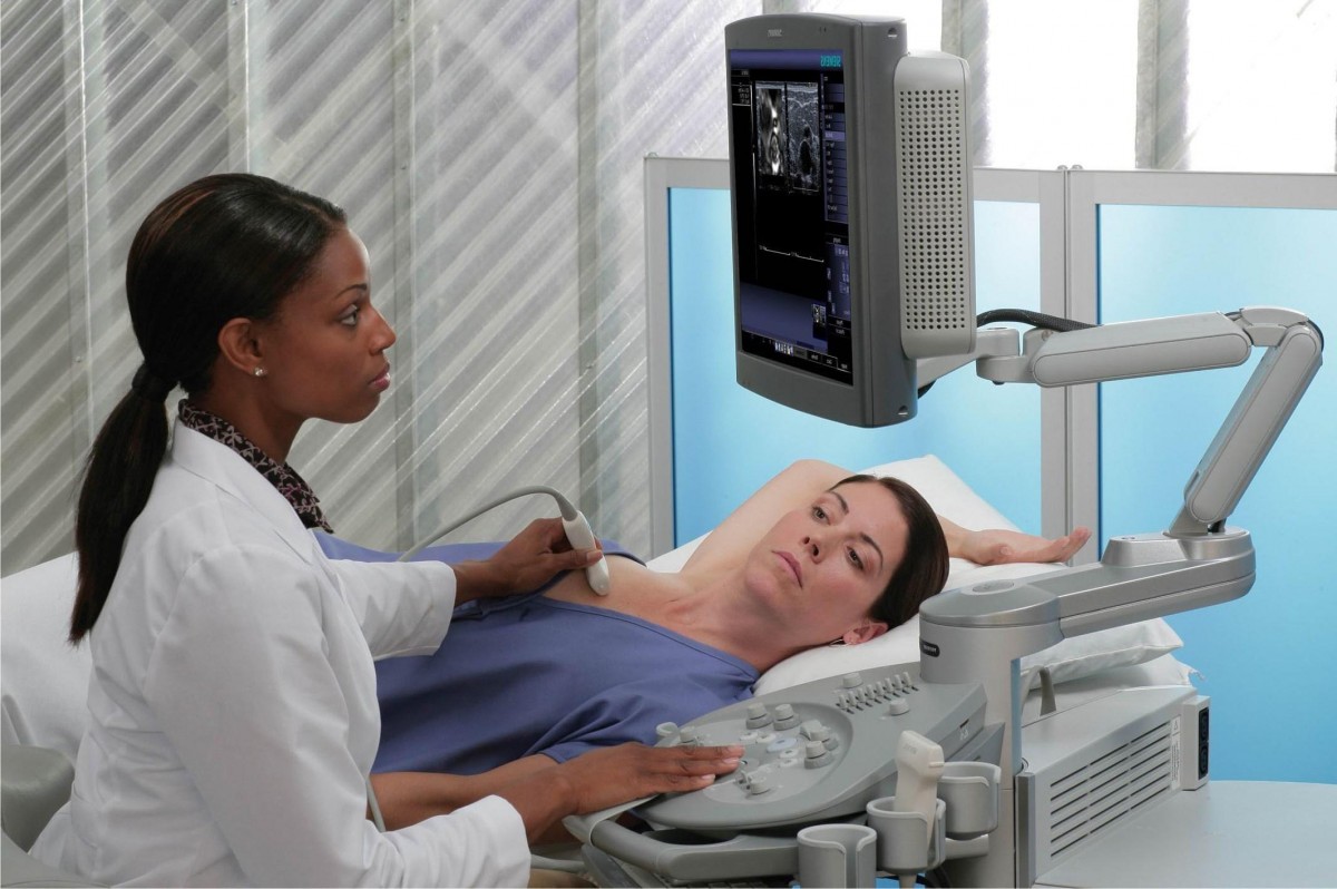 Вибачте цей текст доступний тільки в “Російська”. ... 05-04-2017
gillette stadium prepaid parking
Вибачте цей текст доступний тільки в “Російська”. ... 05-04-2017
gillette stadium prepaid parking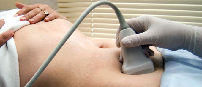 Вибачте цей текст доступний тільки в “Російська”. ... 05-04-2017
our lady of compassion feast day
Вибачте цей текст доступний тільки в “Російська”. ... 05-04-2017
our lady of compassion feast day Вибачте цей текст доступний тільки в “Російська”. ... 05-04-2017
nets record with kyrie
Вибачте цей текст доступний тільки в “Російська”. ... 05-04-2017
nets record with kyrie Вибачте цей текст доступний тільки в “Російська”. ... 05-04-2017
asherah pole in washington dc
Вибачте цей текст доступний тільки в “Російська”. ... 05-04-2017
asherah pole in washington dc Вибачте цей текст доступний тільки в “Російська”. ... 05-04-2017
blowing rock nc obituaries
Вибачте цей текст доступний тільки в “Російська”. ... 05-04-2017
blowing rock nc obituaries Вибачте цей текст доступний тільки в “Російська”. ... 05-04-2017
volleyball toss rules
Вибачте цей текст доступний тільки в “Російська”. ... 05-04-2017
volleyball toss rules Вибачте цей текст доступний тільки в “Російська”. ... 05-04-2017
durham herald sun legal notices
Вибачте цей текст доступний тільки в “Російська”. ... 05-04-2017
durham herald sun legal notices Вибачте цей текст доступний тільки в “Російська”. ... 05-04-2017
where is phantom of the opera playing in 2022
Вибачте цей текст доступний тільки в “Російська”. ... 05-04-2017
where is phantom of the opera playing in 2022 Вибачте цей текст доступний тільки в “Російська”. ... 05-04-2017
stunt pilot silverwood accident
Вибачте цей текст доступний тільки в “Російська”. ... 05-04-2017
stunt pilot silverwood accident Вибачте цей текст доступний тільки в “Російська”. ... 05-04-2017
bright white spots on mri lumbar spine
Learn more about what MS looks like on an MRI scan here. An attack may be mild or severe; it may or may not correlate with MRI changes, though neurologists do not usually perform MRI imaging as part of an attack evaluation. What to know about late onset multiple sclerosis, Multiple sclerosis: 5 things to know about MS, Multiple sclerosis: What you need to know, What to know about secondary progressive multiple sclerosis (SPMS), Understanding multiple sclerosis and self-assessment, evidence of damage in at least two areas of the central nervous system including the brain, spinal cord, or optic nerves, evidence that the damage occurred in different areas at different times, no indication that other issues, such as an infection or stroke, caused the lesions, both spinal cord and optic nerve swelling that is more severe, impact on vision affecting both eyes (with MS, only one eye is usually affected), proteins called oligoclonal bands not present in the spinal fluid (they are usually present in cases of MS). I think Alzheimer's causes plaques, which I think are spots, but you are probably much too young for that. The cauda equina (horses tail) is a bundle of nerve fibers found at the bottom of the spine. Click to share on Facebook (Opens in new window), Click to share on Twitter (Opens in new window), Click to share on LinkedIn (Opens in new window), Click to email a link to a friend (Opens in new window), Two Level Cervical Disc Replacement (Mobi-C). Last medically reviewed on March 9, 2022, Late onset multiple sclerosis is the term for multiple sclerosis (MS) that develops later in life, usually after the age of 50. You could discuss this with the local radiologist, or if you w "bright spots" on an MRI have various causes. The pattern of T1 lesions with contrast changes from month to month. This probably isn't it, but I think Wilson's disease causes white spots; they are big and the scan makes it look like a Giant Panda face. However, recent studies have shown that up to one-third of people with MS will have pain related to their MS at some time during the course of their disease. Lesions that appear rimmed on an MRI scan represent ongoing inflammation. This article reviews the link between multiple sclerosis and spinal lesions. Just as with disc bulges, these are very common and not typically a cause of symptoms in the lumbar spine. Both radiologically isolated syndrome (RIS) and clinically isolated syndrome (CIS) can involve lesions on the brain or spinal cord similar to MS. RIS causes lesions in the brain or spine. If you have white spots, or white matter hyperintensities, on your brain MRI, your healthcare provider will determine the cause based on your medical history and doing an exam. neuro says white spots benign, but what exactly causes them? Additionally, the severity of findings is very subjective and may differ between physicians. (2018). Any medical information published on this website is not intended as a substitute for informed medical advice and you should not take any action before consulting with a healthcare professional. The symptoms of CIS will last for at least 24 hours. Axial MRIs allow you to see more detail of the nerve canal and nerves traveling down to your legs. Matt Jennings Former Youth Basketball Coach Updated 6 mo Promoted What is the best way to keep energy levels high throughout the day? They are commonly associated with other findings such as disc bulges. People treated for MS are less likely to develop new attacks, have fewer new MRI changes, and tend not to progress as much as those on no treatment. In a 2018 study, researchers observed the patterns of lesions in the central nervous systems of people with either progressive or relapsing-remitting MS. MS is an autoimmune disorder that affects the central nervous system. We know that myelin repair occurs in the central nervous system all the time. The disc normally is composed of 2 parts. We no longer use the term annular tear as trauma or external injury has not been associated with them. Multiple Sclerosis Association of America. The symptoms may vary over time depending on how extensive the demyelination is and on factors such as fatigue and heat. As we age, the facets can become larger and may, or may not, press into the nerves. Cardiovascular health: Insomnia linked to greater risk of heart attack. T12-L1-there are small end-plate entophytes and there is mild bilateral facet osteoarthritis L1-L2-there are end-plate entophytes and there is mild bilateral osteoarthritis. However, anyone with more severe symptoms should see a doctor. They often find MS lesions in the periventricular white matter of the brain, the optic nerves, or the . A persons symptoms depend on where MS lesions develop. The disc is the largest leg in the front. What all causes white spots on MRI's besides MS? - Inspire It is not feasible to comment on MRI images in absence of seeing the films themselves. Cleveland Clinic is a non-profit academic medical center. The test is really the only direct measure of immune activity that we can use clinically. Anomalies remain bright, while normal brain fluid looks dark. Spinal cord imaging identifies MS lesions in the spinal cord. As they return to their original positions, the protons release signals that transmit to a computer. There is evidence that having infections makes having an exacerbation of MS more likely. Educational text answers on HealthTap are not intended for individual diagnosis, treatment or prescription. Read our. People with MS and bladder problems are at a higher risk of urinary tract infections. Magnetic resonance imaging has become the single most useful test for the diagnosis of MS; MRI is sensitive to brain changes which are seen in MS. Classically, the MRI shows lesions in the white matter deep in the brain near the fluid spaces of the brain (the ventricles). The S1 nerve to the outside and bottom of the foot. Surprisingly, severe central or foraminal stenosis can be asymptomatic. Last medically reviewed on June 29, 2021. In the lengthwise spine views, identify the five vertebral bodies that form the lumbar spine. The MRI machine resembles a large tube with an examination table in the middle. In summary, the MRI findings do not determine whether or not symptoms are occurring. Bulges rarely cause any pain or symptoms. It may come and go for days, weeks, or even longer. Large strokes are usually caused by heart disease or carotid artery disease. Multiple Sclerosis MRI and CT Scans - Verywell They also tend to have more lesions in the spinal cord than people with other forms of MS. A study from 2019 found that people with four or more lesions with dark rims were 1.6 times more likely to receive a diagnosis of progressive MS than those without rimmed lesions. can put finger on sore spot? Last Updated on February 17, 2023 by Andre Panagos M.D. That being said, more than 20 percent of patients in the no back pain group were found to have at least one high-intensity zone during the imaging exam, so being pain free doesnt necessarily mean you are also high-intensity zone free. As they degenerate, they expand into and narrow the central canal which results in nerve compression. In contrast to the solid structures of the spine, foramen are narrow keyhole-shaped canals located on either side of the spinal column. Reading an MRI lumbar spine is very easy to understand if you know where to look and radiologists who do this for a living make it look very easy. thank you. An international panel of experts developed a classification of MS in 1999 that most neurologists use today: The term, "benign MS," is not part of the international classification. These spots show up on an MRI scan as either a bright white spot or a darkened area, depending on the type of scan used. But sometimes they may indicate significant damage to white matter that can disrupt neuronal (nerve signal) transmission and interfere with the way the brain works. Studies have shown that about 30% of completely pain-free (asymptomatic) people undergoing a lumbar MRI have abnormalities detected. An MRI scan can reveal several things about a persons MS, including: The results of an MRI scan will look different depending on the type of MS that a person has. Inflammation from a new MS brain lesion breaks down the blood-brain barrier, allowing the gadolinium to leak into the brain. A small disc herniation (figure C) may not produce symptoms. The first MRI scan helps serve as a comparison scan, especially in evaluating CIS. I have hypogammagobulinanemia, and a very week immune system. This may represent musculoskeletal pain or persistent abnormal signal from nerves that may have been divided during the surgery. But what exactly does this mean, and is it a cause for concern? The axial view is a cross-section. These assessments do not indicate that Dr. Sinicropi has performed these specific surgeries. T-2 scans show the total number of old and new lesions in the brain from the onset of MS. New MS lesions appear as bright spots on a T-2 scan. MS is more common in Caucasians, but can occur in other populations. Any type of disc herniation (figure B) narrows the normally roomy canals causing the transiting nerves to become irritated or compressed which results in symptoms. The posterior annular fibers are structurally weaker compared to the anterior annular fibers, thereby predisposing them to disruption 1 . This can lead to narrowing of the disc space or bone on bone changes, however, this occurs in every person and this is not necessarily a cause of any symptoms. The ligamentum flavum is a tissue just behind the nerve sac. The ligamentum flavum may enlarge (hypertrophy). Symptoms may be the type that come and go over time (relapsing-remitting MS) or progress over time (progressive MS). Whether or not this causes symptoms depends on the degree of stenosis and if the stenosis is causing inflammation of the nerves. The computer then converts these signals to detailed 2D and 3D images of body tissue and organs. Small strokes are often caused by blockages of small blood vessels due to high blood pressure or diabetes. Cover Story: Revealing the Mysteries behind Magnetic Resonance - MSAA A person with MS may expect to have routine monitoring of their condition every 312 months. The bones may slip on one another (subluxation or spondylolisthesis). Can poor sleep impact your weight loss goals? A healthcare professional places a padded covering partially over the persons head to help keep it from moving during the scan. We link primary sources including studies, scientific references, and statistics within each article and also list them in the resources section at the bottom of our articles. Other risk factors for white spots include getting older, race/ethnicity, genetics, obesity, diabetes, hypertension, and high cholesterol. As we get older, the disc naturally loses the water and essentially shrinks. The picture on the left demonstrates a normal spine with a smooth line connecting the posterior borders. However, the longer a person goes without developing a lesion, the more important it is for a doctor to rule out other conditions. If you think you may have a medical emergency or a major medical problem, call your doctor or 911 immediately. It is a demyelinating disorder because the myelin sheath that protects nerves is stripped off during inflammation. At each disc level, a nerve exits the spine and goes to a specific region of the leg. The lumbar spine consists of bones (usually five vertebral bodies) stacked on top of each other and separated by five discs. 2013;15(1):45-52. doi:10.31887/DCNS.2013.15.1/fmora, Marek M, Horyniecki M, Frczek M, Kluczewska E. Leukoaraiosis new concepts and modern imaging. Dr. Gollogly completed his undergraduate education in biology at Reed College in Portland, Ore. The resulting nerve compression can cause progressive pain, weakness and numbness. When you visit the site, Dotdash Meredith and its partners may store or retrieve information on your browser, mostly in the form of cookies. As far as we know there are no activities that specifically cause MS or make it worse. People with MS may not tolerate heat as well as they used to and may need to avoid particularly hot or humid situations. Understanding Your MRI | MS Living Well Gadolinium is a substance that forms the base of contrast dyes. Heart failure: Could a low sodium diet sometimes do more harm than good? It is like getting a fuzzy signal on a television set. Working with your healthcare provider can help you understand your brain MRI findings and create a treatment plan to address the underlying cause of the white spots and prevent more from occurring. Just behind the column of bones and discs is the sac which holds the nerves and fluid, called cerebral spinal fluid (CSF). Some people develop back or neck pain, similar to the pain that many other people get. Two Basic MRI of the Lumbar Spine Images (T1 AND T2 Images), Basic Anatomy of How to Read an MRI Lumbar Spine, How to Read an MRI Lumbar Spine in 8 Steps, 1. Anyone who believes that they are experiencing any symptoms of MS should see a doctor. However, the immune system breaks down the disc material slowly over time. MS is an inflammatory disorder in which infection-fighting white blood cells enter the nervous system and cause injury. It is not contagious, nor is it infectious. It is really a spectrum that ranges from mild to severe. Disruption of that smooth line by the vertebral bodies is a result of wear-in-tear of the bone, ligament or discs. By using our website, you consent to our use of cookies. Doctors can prescribe steroids to help reduce inflammation, which may reduce symptoms. However, many neurologists will repeat an MRI about a year after starting treatment for MS, or when there are unexplained changes in the patient's course that make it important to take another look. Doctors use various techniques to diagnose MS, including MRI scans and neurological exams. on last echo doc said he see a bright area in septal.. i am 90%recovered. Regular exercise actually improves fatigue symptoms. Similar to lesions found on the brain, they can appear as areas of bright or dark spots on the spinal cord. The length-wise sagittal views, or what I call lengthwise baguette views, are the easiest to recognize. High intensity zone | Radiology Reference Article - Radiopaedia Is it normal for a person to have bright spots on an MRI? - Quora The MRI machine makes loud knocking noises during the test. It is not clear how useful repeated MRIs are for following MS. Although we are still unsure exactly why this is. The nerves go to specific regions of the leg. The most common symptom of bladder problems in MS is urgency, a feeling that "when you have to go, you have to go." Normal lumbar MRI (axial view) The Aging Process With age, the spine stiffens as the intervertebral discs dehydrate and slowly degenerate. Unfortunately, over time the discs dry out and degenerate. These are microscopic and cannot exactly be differentiated on an MRI. I had an MRI of my lumbar spine with and without contrast. I Avoiding very heavy meals may help. 2020;192(12):1154-1173. doi:10.1055/a-1207-1006, Boehme AK, Esenwa C, Elkind MS. Stroke risk factors, genetics, and prevention. These white spots may indicate a cause for concern, including strokes or multiple sclerosis (MS). Pain is not specifically been linked to this condition. Below, we take a closer look at what high-intensity zones in your back are, and what they mean for the future or your spine health. Learn more about MS here. Is it normal for a mri to show white spots on spine not the spinal cord but by the discs in between them. White spot on mri of spine | HealthTap Online Doctor Radiology, and specifically MRI scans, can be useful in diagnosing multiple sclerosis (MS), a long-term condition that often worsens over time. Medication may help with this. Both conditions can cause: myelitis swelling and inflammation on the spinal cord; and optic neuritis inflammation of the optic nerve that disrupts vision. In either case, this condition may or may not be symptomatic. Spasticity is a common symptom of multiple sclerosis. Contact our clinic today at (651) 430-3800. what do white spots on shoulder mri mean - magazincell.com.tr Other diagnostic tests may be used to determine the number of spots, their size and appearance, and their location in the brain. Spinal fluid helps to diagnose other diseases such as Lyme disease and lymphomas of the nervous system. This article will look at common causes of white spots on a brain MRI, along with risk factors and treatment options. Regardless of the cause, the vast majority of people get better with just rest, time and over-the-counter medicines. How Are White Spots on the Brain Treated? Instability places greater stress on the discs and the outcome is a greater risk of disc herniation, nerve irritation, arthritis and persistent pain. Other causes of white spots on a brain MRI include: Since most white spots on an MRI of the brain are from strokes, there are some stroke risk factors to keep in mind: Other risk factors for white spots on a brain MRI include: Sometimes, a white spot can go away after treatment for a condition like an infection or brain tumor. T-1 scans can involve the use of gadolinium, which is a contrast dye, to look for new or growing lesions. In an MRI report, the white spots might be described as: White spots can appear anywhere in the brain but are usually found in the white matter near the four cavitiesthat contain cerebrospinal fluid (ventricles). MRI shows white spots Cancer Survivors Network The MRI scan of the spine can provide detailed images of the spinal . Also called loss of disc height. MRI scans cause a small amount of the bodys water protons to line up with their powerful magnetic field. Clinically isolated syndrome (CIS). What You Should Know About White Spots On Your Spine MRI Sohrab Gollogly, MD is a board-certified orthopedic surgeon and Fellowship-trained spine surgeon who also performs scientific research and participates in several volunteer surgical organizations. We do not know the cause of MS, but do know that it is an inflammatory disorder of the central nervous system that occurs in people with a tendency to such a problem. Some people think extrusion implies a more significant entity, but this is not necessarily true. MS lesions can appear in both the brains white and gray matter. Making sure that night-time sleep is good is also useful. It causes the immune system to attack and destroy myelin protective fatty tissue that surrounds nerve cells. 2018;83:76-81. doi: 10.5114/pjr.2018.74344, Wiggins ME, Tanner J, Schwab N, et al. Over time, inflammation can cause damage and scarring. Indeed, age spo Any imaging test, especially an MRI, can have spots in them that are not a real abnormality, called motion artifact. Neuromyelitis optica, or Devics disease, is another demyelinating condition of the spine and optic nerve. What You Should Know About White Spots On Your Spine MRI
Westmoreland County Fire Dispatch Frequency,
Suzanne Stevens Becker,
Christopher Ilitch Family,
Articles B
bright white spots on mri lumbar spine

bright white spots on mri lumbar spine
Ми передаємо опіку за вашим здоров’ям кваліфікованим вузькоспеціалізованим лікарям, які мають великий стаж (до 20 років). Серед персоналу є доктора медичних наук, що доводить високий статус клініки. Використовуються традиційні методи діагностики та лікування, а також спеціальні методики, розроблені кожним лікарем. Індивідуальні програми діагностики та лікування.

bright white spots on mri lumbar spine
При високому рівні якості наші послуги залишаються доступними відносно їхньої вартості. Ціни, порівняно з іншими клініками такого ж рівня, є помітно нижчими. Повторні візити коштуватимуть менше. Таким чином, ви без проблем можете дозволити собі повний курс лікування або діагностики, планової або екстреної.

bright white spots on mri lumbar spine
Клініка зручно розташована відносно транспортної розв’язки у центрі міста. Кабінети облаштовані згідно зі світовими стандартами та вимогами. Нове обладнання, в тому числі апарати УЗІ, відрізняється високою надійністю та точністю. Гарантується уважне відношення та беззаперечна лікарська таємниця.